NCERT Solutions Class 11 Biology Chapter 18 - Body Fluids and Circulation

Q1: Name the components of the formed elements in the blood and mention one major function of each of them.
Ans: The component elements in the blood are:
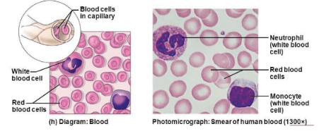
(i) Erythrocytes: They are the most abundant cells and contain the red pigment called haemoglobin. They carry oxygen to all parts of the body. Red blood cells are produced continuously in some parts of the body, such as the marrow of long bones, ribs, etc. There are about 4 – 6 million RBCs per cubic millimetre of blood.
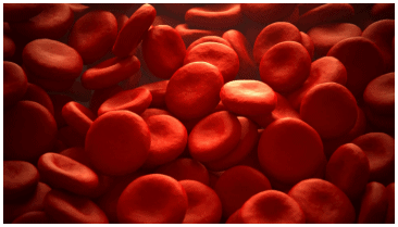 Erythrocytes
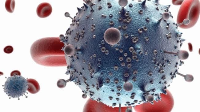
(ii) Leukocytes: Leukocytes are colourless cells. These cells do not contain haemoglobin.
They are the largest cells of the body and are divided into two main categories:
(a) Granulocytes: These leucocytes have granules in their cytoplasm and include neutrophils, eosinophils, and basophils. Neutrophils are phagocytic cells that protect the body against various infecting agents. Eosinophils are associated with allergic reactions, while basophils are involved in inflammatory responses.
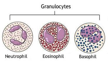 Granulocytes
(b) Agranulocytes: Lymphocytes and monocytes are agranulocytes. Lymphocytes generate immune responses against infecting agents, while monocytes are phagocytic in nature.
(iii) Platelets: Platelets are small irregular bodies present in the blood. They contain essential chemicals that help in clotting. The main function of platelets is to promote clotting.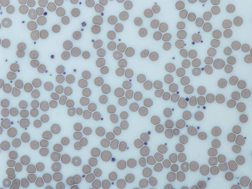 Platelets
Q2: What is the importance of plasma proteins?
Ans: Plasma is the colourless fluid of blood which helps in the transport of food, CO2, waste products, and salts. It constitutes about 55% of blood. About 6.8% of the plasma is constituted by proteins such as fibrinogens, globulins, and albumins.
- Fibrinogen is a plasma glycoprotein synthesised by the liver. It plays a role in the clotting of blood. Globulin is a major protein of the plasma. It protects the body against infecting agents. Albumin is a major protein of the plasma. It helps in maintaining the fluid volume within the vascular space.
 Plasma Protein
Q3: Match column I with column II: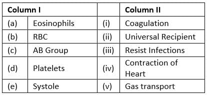
Ans: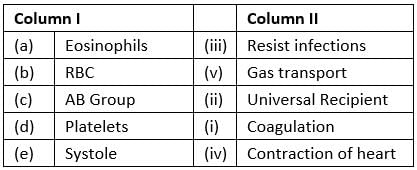
Q4: Why do we consider blood as a connective tissue?
Ans: Connective tissues have cells scattered throughout an extracellular matrix. They connect different body systems.
Blood is considered a type of connective tissue because of two reasons:
 Connective Tissue
- Like the other connective tissues, blood is mesodermal in origin.
- It connects the body systems, transports oxygen and nutrients to all the parts of the body, and removes waste products. Blood has an extra-cellular matrix called plasma, with red blood cells, white blood cells, and platelets floating in it.
Q5: What is the difference between lymph and blood?
Ans: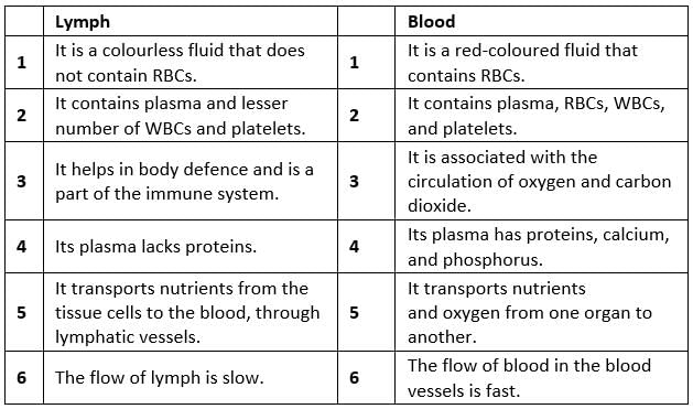
Q6: What is meant by double circulation? What is its significance?
Ans: Double circulation is a process during which blood passes twice through the heart during one complete cycle.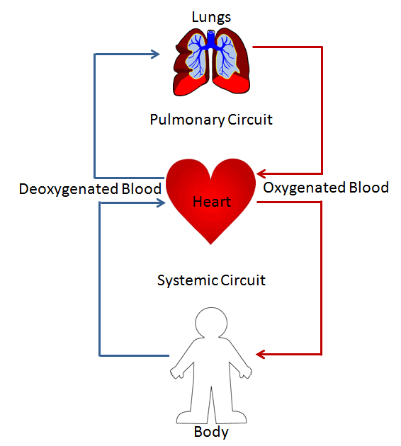 Double Circulation This type of circulation is found in amphibians, reptiles, birds, and mammals. However, it is more prominent in birds and mammals as in them the heart is completely divided into four chambers – the right atrium, the right ventricle, the left atrium, and the left ventricle.
The movement of blood in an organism is divided into two parts:
1. Systemic circulation
2. Pulmonary circulation
- Systemic circulation involves the movement of oxygenated blood from the left ventricle of the heart to the aorta. The blood then carries it through a network of arteries, arterioles, and capillaries to the tissues. The venules, veins collect the deoxygenated blood, and vena cava from the tissues and is emptied into the left auricle.
- Pulmonary circulation involves the movement of deoxygenated blood from the right ventricle to the pulmonary artery, which then carries blood to the lungs for oxygenation. From the lungs, the oxygenated blood is carried by the pulmonary veins into the left atrium.
Hence, in double circulation, blood has to pass alternately through the lungs and the tissues.
Significance of double circulation: The separation of oxygenated and deoxygenated blood allows a more efficient supply of oxygen to the body cells. Blood is circulated to the body tissues through systemic circulation and to the lungs through the pulmonary circulation.
Q7: Write the differences between:
(a) Blood and Lymph
(b) Open and Closed system of circulation
(c) Systole and Diastole
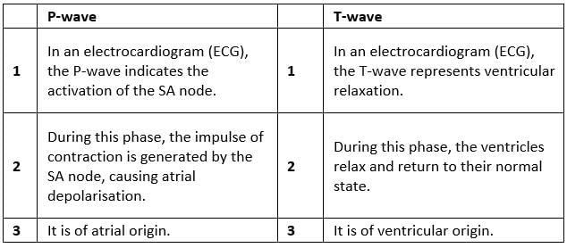
(d) P-wave and T-wave
Ans: (a) Blood and lymph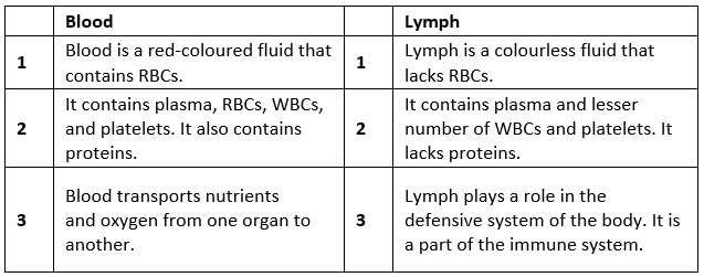 (b) Open and closed systems of circulation
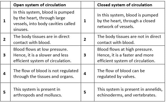 (c) Systole and diastole
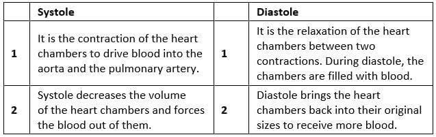 (d) P-wave and T-wave
Q8: Describe the evolutionary change in the pattern of heart among the vertebrates.
Ans: The pattern of the heart has evolved significantly among vertebrates, reflecting increasing complexity:
- Fishes:
- Have a 2-chambered heart with one atrium and one ventricle.
- Pumps deoxygenated blood to the gills for oxygenation.
- Blood flows in a single circulation system.
- Amphibians and Reptiles:
- Possess a 3-chambered heart with two atria and one ventricle.
- Oxygenated blood from the lungs or gills enters the left atrium.
- Deoxygenated blood from the body enters the right atrium.
- Blood mixes in the single ventricle, leading to incomplete double circulation.
- Crocodiles, Birds, and Mammals:
- Have a 4-chambered heart with two atria and two ventricles.
- Allows complete separation of oxygenated and deoxygenated blood.
- Enables double circulation, where blood flows through two separate pathways without mixing.
Q9: Why do we call our heart myogenic?
Ans: In the human heart, contraction is initiated by a specially modified heart muscle known as sinoatrial node. It is located in the right atrium. The SA node has the inherent power of generating a wave of contraction and controlling the heartbeat. Hence, it is known as the pacemaker. Since the SA node initiates the heartbeat and the impulse of contraction originates in the heart itself, the human heart is termed myogenic. The hearts of vertebrates and molluscs are also myogenic.
Q10: Sino-atrial node is called the pacemaker of our heart. Why?
Ans: The sino-atrial (SA) node is a specialised group of cells located in the upper part of the heart's right atrium. It serves as the heart's natural pacemaker for the following reasons:
- The SA node generates electrical impulses that initiate the heartbeat.
- These impulses trigger a series of electrical events in the heart, leading to muscle contractions.
- It controls the rhythm of the heart, ensuring it beats in a coordinated manner.
- The SA node typically produces 70-75 impulses per minute, matching the average heart rate.
Due to its role in starting and regulating the heart's rhythm, the SA node is essential for effective blood pumping throughout the body.
Q11: What is the significance of atrio-ventricular node and atrio-ventricular bundle in the functioning of heart?
Ans: The atrioventricular (AV) node is present in the right atrium, near the base of the interauricular septum that separates the right auricle from the ventricle. It gives rise to the bundle of His that conducts the cardiac impulses from the auricles to the ventricles.
- As the bundle of His passes the ventricle along the inter-ventricular septum, it divides into two branches – the right ventricle and the left ventricle. The end branches of this conducting system then form a network of Purkinje fibres that penetrate the myocardium.
- The auricular contraction initiated by the wave of excitation from the sinoatrial node (SA node) stimulates the atrio-ventricular node, thereby leading to ventricles' contraction through the bundle of His and Purkinje fibres. Hence, the atrioventricular node and the atrioventricular bundle play a role in the contraction of ventricles.
Q12: Define a cardiac cycle and cardiac output.
Ans: The cardiac cycle refers to the series of events that occur in the heart from the start of one heartbeat to the next. It includes:
- Atrial systole: Contraction of the atria.
- Ventricular systole: Contraction of the ventricles.
- Cardiac diastole: Relaxation phase of the heart.
The heart typically completes about 72 cycles per minute, with each cycle lasting approximately 0.8 seconds. During each cycle, each ventricle pumps around 70 mL of blood, known as the stroke volume.
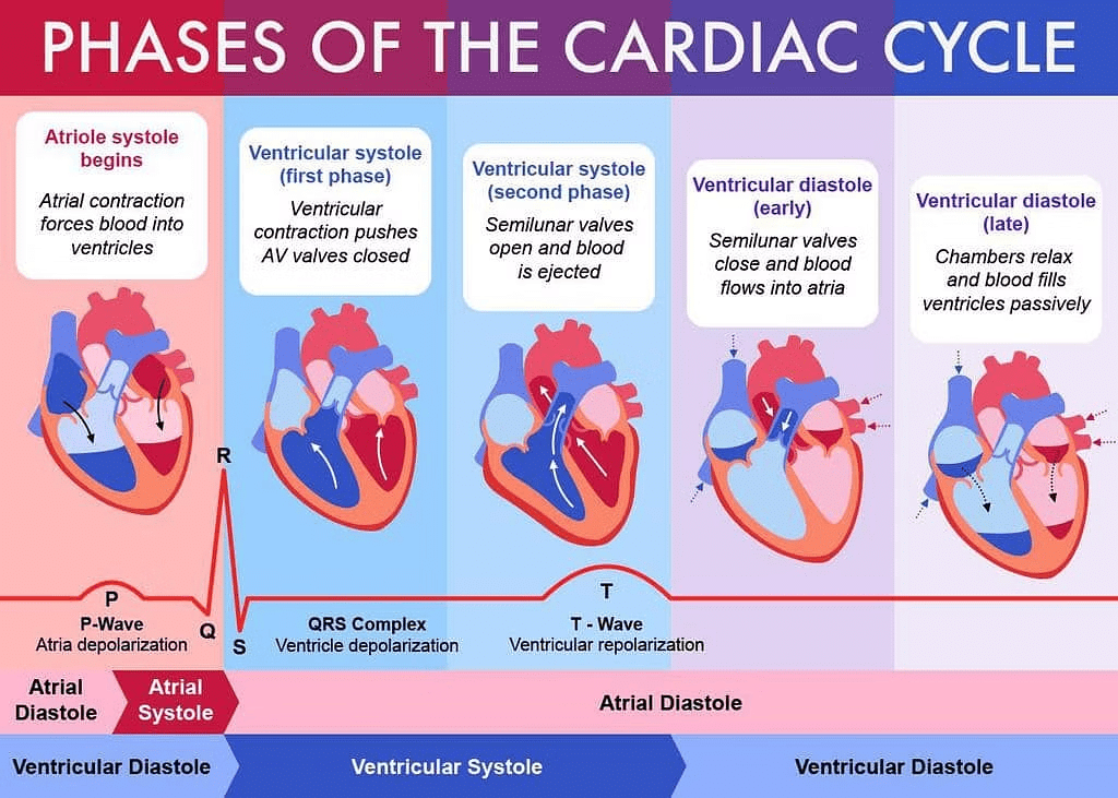
Cardiac output is the total volume of blood ejected by the ventricles in one minute. It is calculated by multiplying the stroke volume by the heart rate, averaging about 5 litres in a healthy adult.
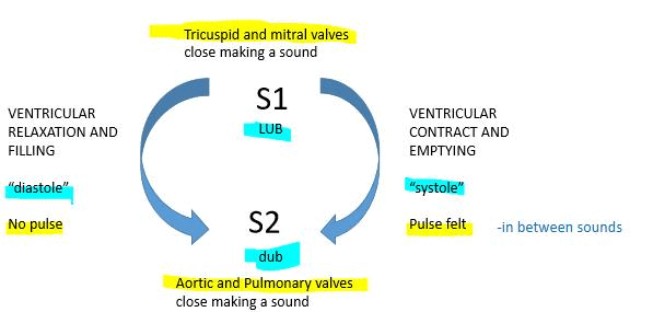
Q13: Explain heart sounds.
Ans: Heart sounds are the noises produced by the heart as it functions. In a healthy heart, there are two main sounds known as lub and dub.
- Lub: This is the first heart sound, occurring when the tricuspid and bicuspid valves close at the start of systole.
- Dub: This is the second heart sound, which happens when the semilunar valves close at the beginning of diastole.
These sounds are crucial as they provide insights into the heart's condition and functioning.
 Heart Sounds
Q14: Draw a standard ECG and explain the different segments in it.
Ans: An electrocardiogram is a graphical representation of the cardiac cycle produced by an electrograph.
The diagrammatic representation of a standard ECG is shown below:
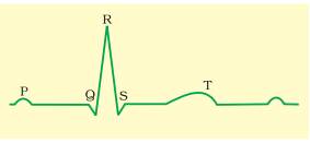 ECG
ECG
The P-wave signifies the electrical excitation (depolarization) of the atria, causing both atria to contract.
The QRS complex shows the depolarization of the ventricles, triggering their contraction, which begins shortly after the Q wave and marks the onset of systole.
The T-wave represents the repolarization of the ventricles, as they return to their normal state, and its end signals the completion of systole.
By counting the number of QRS complexes within a certain time period, the heart rate of an individual can be determined.
|
150 videos|399 docs|136 tests
|
FAQs on NCERT Solutions Class 11 Biology Chapter 18 - Body Fluids and Circulation
| 1. What are the main components of blood and their functions? |  |
| 2. How does the circulatory system maintain homeostasis? | |
| 3. What is the difference between arteries, veins, and capillaries? | |
| 4. What role does the heart play in circulation? | |
| 5. How does the lymphatic system contribute to the circulatory system? | |


practice quizzes
,NCERT Solutions Class 11 Biology Chapter 18 - Body Fluids and Circulation
,Free
,Sample Paper
,Semester Notes
,shortcuts and tricks
,Exam
,Viva Questions
,mock tests for examination
,past year papers
,video lectures
,ppt
,Important questions
,study material
,Summary
,MCQs
,NCERT Solutions Class 11 Biology Chapter 18 - Body Fluids and Circulation
,Objective type Questions
,NCERT Solutions Class 11 Biology Chapter 18 - Body Fluids and Circulation
,Previous Year Questions with Solutions
,Extra Questions
;


NCERT Solutions: Body Fluids & Circulation Free PDF Download
Importance of NCERT Solutions: Body Fluids & Circulation
NCERT Solutions: Body Fluids & Circulation Notes
NCERT Solutions: Body Fluids & Circulation NEET Questions
Study NCERT Solutions: Body Fluids & Circulation on the App
|
© EduRev
|
Education Revolution
|


|


