NEET Exam  >  NEET Notes  >  Biology Class 11  >  The Human Brain

The Human Brain | Biology Class 11 - NEET PDF Download

Nervous System

  • System which regulate the various activities of the body through nerve impulses is called the nervous system. Through this system the messages are transmitted at a faster rate.
  • The nervous – system controls and also co-ordinates the various activities of the organs of the animals.
  • Whole nervous – system of human being is derived from embryonic Ectoderm.

 The Human Brain | Biology Class 11 - NEET

         The Human Brain | Biology Class 11 - NEET

Central Nervous System

It includes the brain and the spinal-cord. These are formed from the neural-tube which develops from the ectoderm after the gastrula stage of embryo.

Development of CNS :– It develops from neural tube in intrauterine life (I.U.L.). Anterior part of neural tube develops into brain while caudal part of neural tube develops into spinal cord.

Brain's approximately 70-80% part of brain develops in 2 year of age & complete development is achieved in 6 years of age & spinal cord develops completely in 4 to 5 years of age.

Brain

It is situated in cranial box which is made up of 1 frontal bone, 2 parietal bone, 2 temporal bone, 1 occipital bone. The weight of brain of an adult man is 1400 gm and of female is 1250 gm.

Brain Meninges

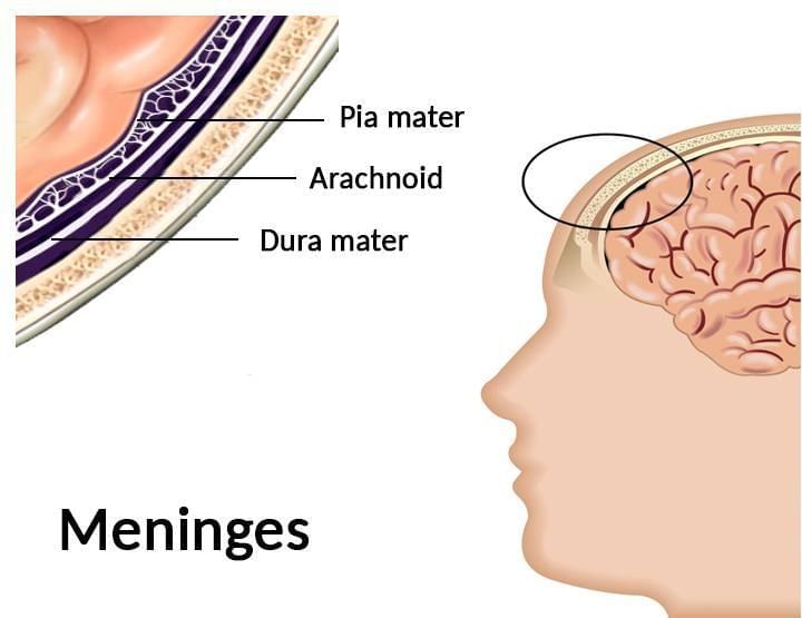
Brain is covered by three membranes of connective tissue termed as meninges or menix.

The Human Brain | Biology Class 11 - NEET

(1) Dura Mater

  • This is the first and the outermost membrane which is thick, very strong and non- elastic. 
  • It is made up of collagen fibres. 
  • This membrane is attached with the innermost surface of the cranium.
  • It is double layer–outer Endosteal layer which is closely attached with inner most surface of cranium & no space is found between skull & Dura mater (No Epidural space). 
  • Inner meningeal layer which is related with other meninges of brain, both are vascular
  • Generally both layers are fused with each other, but at some places these are separated from one another & form a sinus called cranial venous sinus. These sinuses are filled with venous blood.

(2) Arachnoid

  • It is middle, thin and delicate membrane, made up of connective tissue. 
  • It is found only in mammals.
  • It is non vascular layer. Infront of cranial venous sinus, it becomes folded, these folds called Arachnoid villi.
  • These villi reabsorb the cerebrospinal fluid (CSF) from subarachnoid space & pour it into cranial venous sinuses. 

(3) Piamater

  • It is Inner most, thin and transparent membrane, made up of connective tissue.
  • Dense network of blood capillaries are found in it, so it is highly vascular.
  • It is firmly adhere to the brain. Piamater & arachnoid layer at some places fuse together to form leptomeninges.
  • Piamater merges into sulci of brain & densely adhere to it. At some places it directly merges in the brain and called telachoroidea Tela choroidea form the choroid plexus in the ventricles of brain.

Sub Dural Space :– Space between duramater & arachnoid. It is filled with serous fluid.

Sub Arachnoid Space :– Space between Arachnoid & Piamater is filled with C.S.F. Cranial nerves also pass through this space

 Meningitis : Any inflammation of menix is called as meningitis. It may be caused by viruses, bacteria or protozoa.

Cerebrospinal Fluid (CSF) 

  • This fluid is clear and alkaline in nature just like lymph. It has protein (Albumin, globulin), glucose, cholesterol, urea, bicarbonates, sulphates and chlorides of Na, K. Protein & cholesterol concentration is lesser than plasma & Cl– conc. is higher than plasma. 
  • In a healthy man, in 24 hrs, 500 ml of C.S.F is formed & absorbed by arachnoid villi. At a time total volume of C.S.F. is 150 ml.
  • CSF is present in ventricle of brain, subarachnoid space of brain & spinal cord.
  • Formation :- Mainly in choroid plexus of lateral ventricles, minor quantity is formed in IIIrd ventricle & IVth ventricle.
  • Collection of CSF for any investigation is done by lumbar puncture (LP). It is done at L3–L4 region.
  • Spinal anaesthesia is also given by L.P.

Functions of CSF

  • Protection of Brain :- It act as shock absorbing medium and work as cushion.
  • It provides buoyancy to the brain
  • Excretion of waste products.
  • Endocrine medium for the brain to transport hormones to different areas of the brain.

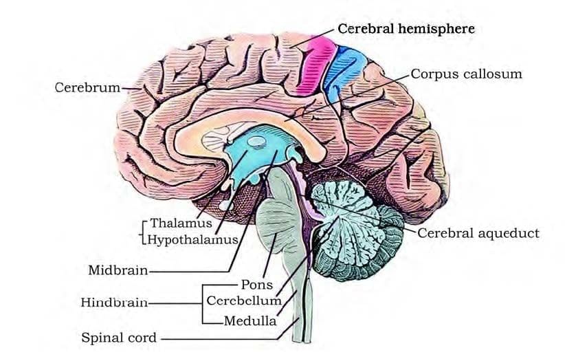
Human brain divided into three parts – 

(1) Fore brain – Cerebrum, Diencephalon.

(2) Mid brain – Optic lobes and Crura cerebrai.

(3) Hind Brain – Pons, Cerebellum, Medulla During embryonic stage, brain develops from three hollow vesicles –

The Human Brain | Biology Class 11 - NEET

Cerebrum

The Human Brain | Biology Class 11 - NEET

  • It is first & most developed part of brain.
  • It makes 2/3 part of total brain
  • Cerebrum consist of two cerebral hemispheres on the dorsal surface. A longitudinal groove is present between two cerebral hemispheres called as median fissure. Both the cerebral hemispheres partially connected with each other by curved thick nerve fibres called corpus callosum.
  • Each cerebral hemisphere is divided into 5-lobes – Anterior, middle, posterior, lateral and insula lobe.
  • Anterior lobe is also called frontal lobe (largest lobe). Middle lobe is also called parietal lobe.
  • Central sulcus separates frontal lobe from parietal lobe. Lateral lobe or temporal lobe is separated from frontal lobe & parietal lobe by incomplete sulcus called lateral sulcus.
  • Posterior lobe is also called occipital lobe, it is separated from parietal lobe by a sulcus called parieto occipital sulcus.
  • In right handed person, left hemisphere is dominant while in left handed person right hemisphere is dominant.
  • Many ridges & grooves are found on dorsal surface of cerebral hemisphere. Ridges are known as gyri while grooves are called sulci. These cover the 2/3 part of cerebrum.
  • Gyri & sulci are more developed in human being so human being are most intelligent living beings.

The Human Brain | Biology Class 11 - NEET

Corpus Callosum

  • It is largest commissure of Brain.
  • Exclusive feature of mammals.Curved thick band of white nerve fibre situated between two cerebral hemisphere in the median fissure. Anterior truncated part of corpus callosum is called Genu while posterior truncated part is called splenium.
  • A oblique band is formed by body of corpus callosum & it goes towards Genu called fornix.
  • A small cavity is developed among body of callosum, Genu & fornix called as Vth ventricle or pseudocoelom. This ventricle is covered by a thin membrane called as septum pellucidum.

The Human Brain | Biology Class 11 - NEET

Diencephalon :– It is small and posterior part of fore brain. It is covered by cerebrum. It consists of Thalamus, hypothalamus, Epithalamus & Metathalamus. 

(i) Thalamus : It forms upper lateral walls of Diencephalon. It forms 80% part of Diencephalon. Gate keeper of brain.

It acts as a relay centre. It receives all sensory impulses from all part of body & these impulses are send to the cerebral cortex.

(ii) Hypothalamus : It forms lower or ventral part of Diencephalon.

A cross like structure is found on anterior surface of hypothalamus called as optic chiasma.

Pituitary body is attached to middle part of hypothalamus by infundibulum.

Corpus mamillare or Corpus albicans or mammillary body is found on the posterior part of hypothalamus. It is a character of mammalian brain.

Epithalamus – It forms the roof of diencephalon. Pineal body (Epiphysis cerebri) is found in epithalamus.

Metathalamus – It consists of medial geniculate body & lateral geniculate body. It is located in the floor of Diencephalon.

Mid brain – It is small & contracted part of brain. Anterior part of mid brain contains two longitudinal myelinated nerve fibres peduncles called Cerebral peduncles or crus cerebri or crura cerebri. On the posterior part of mid brain are found four spherical projection called colliculus or optic lobes. Four colliculus are collectively called as corpora  quadrigemina (2 upper & 2 lower)..

Only 2 colliculus or optic lobes are found in mid brain of frog called as corpora bigemina.

The Human Brain | Biology Class 11 - NEET

Hind Brain : 3 Parts –

(1) Pons (2) Cerebellum (3) Medulla Oblongata (M.O.)

(1) Pons or Pons varolii : It is small spherical projection, which is situated below the midbrain & upper side of the M.O. It consists of many transverse & longitudinal nerve fibres. Transverse nerve fibres connect with cerebellum (Lateral lobes of cerebellum).

Longitudinal fibres connect cerebrum to M.O.

(2) Cerebellum : Made up of 3 lobe [2 lateral lobe & 1 vermis (divide in 9 segments)].

Both lateral lobes become enlarged & spherical in shape, so lateral lobe of cerebellum are also called as cerebellar hemisphere. Due to this reason, regulation & Coordination of voluntary muscle is more developed as compared to other animals.

In terminal part of vermis, one pair of flocculonodular lobes are found. These continue in the form of flocculus. Three cerebellar peduncle are formed, superior cerebellar peduncle attach with mid brain.

Middle cerebellar peduncle attach with pons and Inferior cerebellar peduncle attach with M.O.

The Human Brain | Biology Class 11 - NEET

(3) Medulla Oblongata (M.O.) : Posterior part of brain  is tubular & cylindrical in shape.

Mid brain, pons & medulla are situated in one axis and called as Brain stem.

Internal Structure of Brain – One pair of olfactory lobes are small spherical & solid in human brain. No ventricle is found in it. Both olfactory lobe are separate with each other & are embedded into ventral surface of the both Frontal lobe of cerebral Hemisphere. Olfactory centre is situated in temporal lobe.

 The Human Brain | Biology Class 11 - NEET

The Human Brain | Biology Class 11 - NEET


  • Except Midbrain, Cerebellum, Pons & olfactory lobe complete brain is internally hollow. Its cavity is lined by ependymal epithelium. (Ciliated Columnar Epithelium)
  • Cavities of brain are known as ventricles, filled with cerebrospinal fluid (C.S.F.).  
  • In Rabbit, Cavity of olfactory lobe is hollow called as 1st ventricle or Rhinocoel. Both Rhinocoel continue in cavity of cerbral hemisphere, known as 2nd ventricle or paracoel or lateral ventricle.
  • In human 1st and 2nd ventricles are considered as paracoel or Lateral ventricle.
  • On the posterior side, both paracoel combine with each other & open into cavity of Diencephalon through an aperture known as Foramen of Monro.
  • Cavity of diencephalon is known as 3rd ventricle or Diocoel.
  • A tent shape space or cavity present between anterior pons, medulla & posterior cerebellum called IVth ventricle
  • 3rd and 4th ventricle are connected with each other through a hollow tube known as Iter or Aqueduct of sylvius. IVth ventricle continues in the metacoel and metacoel continues in the cavity of spinal cord called neurocoel or central canal.
  •  One aperture is found on dorsal surface of metacoel known as foramen of Magendie. Two apertures are found on lateral sides of metacoel know as Foramen of Luschka.[1–1]
  •  The CSF of brain comes out from the foramen of Magendie & Luschka & is poured into sub-arachnoid space.

Histology of Brain

The Human Brain | Biology Class 11 - NEET

  • On dorsal surface of cerebral hemisphere, gray matter becomes more thick, this thick layer of gray matter is known as Cerebral cortex/Neopallium/Pallium.
  • Outer part of cerebellum is made up of gray matter while inner part is of white matter. White matter projects outside & forms a branched tree like structure known as Arbor Vitae.

Choroid plexus : Tela choroidea (Pia mater which is merge in ventricle)  +

Blood capillaries + Ependymal epithelium Site : Two major plexuses in lateral ventricles. 2 minor plexuses in IIIrd ventricle 1 minor plexus in IVth ventricle.

Function : Formation of CSF by secretion of plasma.

Some time (Congenitally or Infection) Iter or aqueduct become blocked therefore improper circulation of CSF or blockage of CSF circulation occur therefore Intra cranial pressure increases, head becomes enlarged, this condition called Hydrocephalus.

Circulation :- From the ventricles CSF comes into subarachnoid space through foramen of Magendie & foramen of Luschka.

In sub arachnoid space, CSF  is absorbed by arachnoid villi which pour it into cranial venous sinus. From venous sinus CSF enters in blood circulation.


 The Human Brain | Biology Class 11 - NEET

The Human Brain | Biology Class 11 - NEET


Basal Nuclei/ Basal ganglie : 

Situated in the wall of cerebral hemisphere.

The Human Brain | Biology Class 11 - NEET

The Human Brain | Biology Class 11 - NEET


Function : 

(1) It maintains muscle tone.

(2) It regulates automatic associated movement like swinging of arms during walking

(3) In lower animals, when cerebral cortex is not developed basal nuclei acts as motor centre.

Lesion in basal nuclei leads to a disease called as parkinsonism (Rigidity, Tremor at rest, mask like face)

(4) Regulate stereotypic movements.

(5) Releated to initiation & termination of movements.


Reticular activating System :– It is special sensory fibre which is situated in Brain stem & further go into Thalamus. It is related with consiousness, alertness & awakening. Therefore it is also called gate keeper of consiousness.

Limbic System:– It is visible like a wish bone, tuning fork or liplike.

Limbic lobe (area of temporal Lobe) +

Hippocampus + Hypothalamus including septum + Part of Thalamus + Mammalary bodies + Amygdaloid complex


The Human Brain | Biology Class 11 - NEET


Functions of Limbic System : 

(1) Behaviour, Emotion, rage and anger (Hypothalamus, amygdaloid body)

(2) Recent memory & short term memory converts into long term memory. (Hippocampal lobe)

(3) Food habit

(4) Sexual Behaviour

(5) Olfaction (Hypothalamus) (Hypothalamus) (Hippocampal lobe and Limbic lobe)


S.No.

Name of Area

Location

Relation or Analysis

1.

Prefrontal cortex

Eontal lobe

Seat of intelligence, knowledge, creative ideas, ability to abstract, memory, (organ of mind).

2.

Premotor area

Eontal lobe

  • Written centre
  • Associated Movement of eye, head & body.
  • Control complex movement of jaw, tongue, pharynx, larynx

3.

Motor area

Eontal lobe

Analysis of all type of voluntary muscle

4.

Eontal Eye field

Eontal lobe

Responsible for conjugate movement of eye. Opening & closing of eye lid

5.

Broca' s Area

In Ft handed person

In Lt handed person

Erontal lobe present of Lt. side present of Rt. side (Motor speech area)

Analysis for speak if injury to this region inability to speak (aphasia) even though muscle concerned are not paralysed

6.

Auditory area

Temporal

Analysis for sound.

7.

Olfactory

Temporal or Hippocampal gyrus

Analysis for smell.

8.

Wernicke' s area (Sensory area of speech)

Temporal

Analysis for language ,Sensory analysis for speech

9.

Gustatory area

Earidal

Analysis for Taste

10.

Somesthetic area

Earidal

Analysis for touch, pressure, pain, knowledge about position in space. taking in information from environment etc.

11.

Angular gyrus

Earidal

Sansory analysis for writing

12.

Occipital area

Occipital

Analysis for vision


Special point : 

Association area responsible for complex functions like intersensory association, memory and communications.

FUNCTIONS OF BRAIN

1. Olfactory lobe :– It is supposed to be centre of smelling power. Its size is small in mammals comparatively because most of its parts become a part of cerebrum (including olfactory tract) some animals like sharks and dogs have well developed olfactory lobes.

2. Cerebral hemispheres :– It is the most developed part in mammals. It is the most important part of brain because it controls and regulates different parts of brain. This is the centre of conscious senses, will power, voluntary movements, knowledge, memory, speech and thinking, reasoning etc.

Different sense organs send impulses here, and in this part of brain analysis and coordination of impulses is done then messages are transferred according to the reactions through voluntary muscles.

All the voluntary actions are controlled by cerebral hemispheres. 


3. Diencephalon :– Its dorsal side is called epithalamus in which pineal body is situated, that controls the sexual maturity of animal.

  • Thalamus – Act as relay centre for sensory stimulation. In lower animal, Cerebral cortex is not developed then Thalamus act as sensory centre.

It is related with RAS.

It is also act as limbic part

  • Functions of Hypothalamus – 

(1) Thermoregulation

(2) Behaviour and emotion

(3) Endocrine control

 (4) Biological clock system

(5) ANS control.

These are found centres of animal feelings/ Emotions like sleep, anger, intercourse, hate, love, affection, and temperature control emotional, pain, hunger, thirst and satisfaction in the hypothalamus.

  • The regulatory hormones of hypothalamus control the activity of endocrine glands. Modern scientists praposed that hypothalamus is the "master gland" not the pituitary.
  • Optic chiasma found in the hypothalamus carry optic impulses received from eyes to the cerebral hemispheres.

Animal becomes blind if this part is destroyed by chance.

Metathalamus – It is related with MGB & LGB.  MGB related with hearing & LGB related with vision. Nerve fibre of concerning place go through Metathalamus.


4. Mid - Brain :– 

  • Four optic lobes or colliculus present, superior optic lobes are the main centres of pupillary light reflexes.
  • Inferior optic lobes are related acoustic (sound) reflex action.
  • Crura cerebri controls the muscles of limbs.

5. Cerebellum:- By this portion of hind brain, impulses are received from different voluntary muscles, joints and then controlling of the their movements. Their regulation and coordination is the main function of this part of brain i.e. cerebellum maintains the body balance of persons which take alcohol in excess, their cerebellum gets affected,as a result of that they can not maintain their balance and their walking is disturbed.


Thus It is related with fine & skillful voluntary movement & also related with body balance, equillibrium,  posture & tone 


6. Pons :- It regulates the breathing reaction through pneumotaxic centre.

7. Medulla Oblongata:- It is the most important part of brain which controls all the involuntary activities of the body e.g. heart beats, respiration , metabolism, secretory actions of different cells rate of engulfing food etc.

Except this it acts as conduction path for all impulses between spinal cord and remaining portions of brain.

It is also concerned with Reflex– Sneezing reflex , salivation reflex, coughing reflex, swallowing reflex, vomiting reflex, yawning reflex.

The document The Human Brain | Biology Class 11 - NEET is a part of the NEET Course Biology Class 11.
All you need of NEET at this link: NEET
182 videos|365 docs|153 tests

Top Courses for NEET

FAQs on The Human Brain - Biology Class 11 - NEET

1. What is the average weight of the human brain?
Ans. The average weight of the human brain is approximately 1.4 kilograms or 3 pounds. However, the weight can vary depending on factors such as age, gender, and overall health.
2. How many neurons are present in the human brain?
Ans. The human brain is estimated to contain around 86 billion neurons. These neurons are responsible for transmitting information and enabling various functions of the brain.
3. Can the human brain regenerate or grow new neurons?
Ans. Yes, the human brain has the ability to regenerate and grow new neurons, a process known as neurogenesis. It primarily occurs in specific regions of the brain, such as the hippocampus, which is involved in memory and learning.
4. What is the role of the frontal lobe in the human brain?
Ans. The frontal lobe plays a crucial role in various cognitive functions, including decision-making, problem-solving, personality, behavior, and voluntary movement. It is responsible for higher-order thinking and executive functions.
5. Can the human brain multitask effectively?
Ans. While the human brain has the ability to switch between tasks rapidly, it is not efficient at multitasking. Research suggests that focusing on one task at a time leads to better productivity and reduces errors compared to attempting to multitask.
182 videos|365 docs|153 tests
Download as PDF
Explore Courses for NEET exam

Top Courses for NEET

Signup for Free!
Signup to see your scores go up within 7 days! Learn & Practice with 1000+ FREE Notes, Videos & Tests.
10M+ students study on EduRev
Related Searches

The Human Brain | Biology Class 11 - NEET

,

mock tests for examination

,

MCQs

,

Previous Year Questions with Solutions

,

Free

,

Objective type Questions

,

ppt

,

Exam

,

Sample Paper

,

practice quizzes

,

Important questions

,

shortcuts and tricks

,

Extra Questions

,

Viva Questions

,

video lectures

,

study material

,

The Human Brain | Biology Class 11 - NEET

,

The Human Brain | Biology Class 11 - NEET

,

past year papers

,

Semester Notes

,

Summary

,

pdf

;