The Human Eye | Science Class 10 PDF Download

THE HUMAN EYE & COLOURFUL WORLD
Our eye is the most important natural optical instrument. The eye resembles a camera in many ways. It has nearly a spherical shape.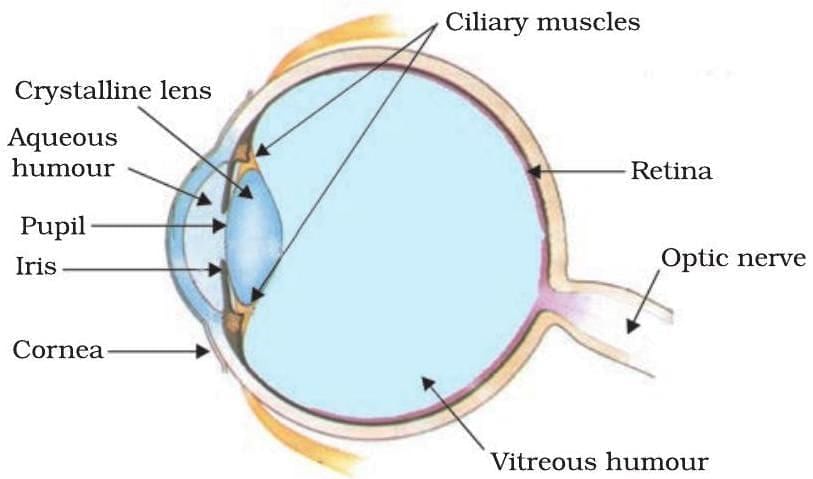
Parts of the Eye and Their Functions
The essential parts of the eye are shown in Fig. and are described below:
(a) Cornea:
- The cornea is a transparent, spherical membrane that covers the front of the eye.
- It allows light to enter the eye.
- Behind the cornea is the aqueous humor, a clear liquid that fills the space.
(b) Iris and Pupil:
- The iris and pupil work together to control the amount of light entering the eye.
- The iris is a dark-colored muscle with a small opening in the center called the pupil.
The iris adjusts the size of the pupil to regulate light intake:
- (i) In bright light, the iris constricts the pupil to reduce light entry.
- (ii) In dim light, the iris expands the pupil to allow more light in.
(c) Eye Lens:
- The eye lens is a convex lens made of transparent, jelly-like protein.
- It is firmer in the center and softer towards the edges.
- The lens is held in place by ciliary muscles, which can change its curvature.
(d) Retina:
- The retina is the inner surface at the back of the eyeball where light is focused.
- It contains about 125 million light-sensitive receptors called rods and cones.
- When light hits these receptors, they send electrical signals to the brain via the optic nerve.
- The space between the lens and retina is filled with a liquid called vitreous humor.
- The image on the retina is held for about 1/16th of a second. This is called persistence of vision.
(e) Blind Spot:
- The blind spot is where the optic nerve leaves the eye to go to the brain.
- There are no rods or cones in this area.
- If an image forms here, it is not seen because no signal is sent to the brain.
Working of the Eye
Light from an object enters the eye through the cornea and the pupil. The eye lens then converges these light rays to form a real, inverted, and smaller image on the retina. The retina contains many light-sensitive cells (cones and rods).
When light hits these cells, they become active and generate electrical signals. These signals are sent to the brain via the optic nerve, allowing us to see the object in its actual size and orientation.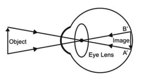
Far Point, Near Point, and Least Distance of Distinct Vision
(a) Far Point:
- The far point is the maximum distance at which the eye can see objects clearly.
- For a normal eye, this point is considered to be at infinity.
(b) Near Point:
- The near point is the closest distance at which the eye can focus on an object clearly.
- For a healthy adult eye, the near point is typically about 25 centimeters from the eye.
(c) Least Distance of Distinct Vision:
- The least distance of distinct vision, often denoted as d or D, is the minimum distance at which the eye can see an object clearly without straining.
- For a normal adult eye, this distance is approximately 25 centimeters.
- This distance tends to increase with age.
Accommodation of the Eye
Accommodation is the property of the eye lens to change its focal length to focus images of objects at varying distances onto the retina. This adjustment is achieved by altering the lens's thickness using the ciliary muscles.
The human eye has the remarkable ability to see both distant and nearby objects clearly. For clear vision, the image of an object must form precisely on the retina. Since the distance between the retina and the eye lens is fixed, the image distance (v) remains constant. However, the object distance (u) varies depending on whether the object is near or far. To maintain a sharp image on the retina, the eye lens adjusts its focal length accordingly.
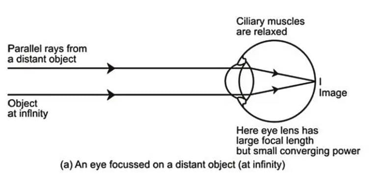
Focusing on Distant Objects
When the eye focuses on distant objects (objects at infinity), the ciliary muscle is fully relaxed. In this state:
The eye lens has minimal thickness.
The focal length is at its maximum, equal to the distance between the eye lens and the retina.
Parallel rays from distant objects are focused precisely on the retina, resulting in clear vision.
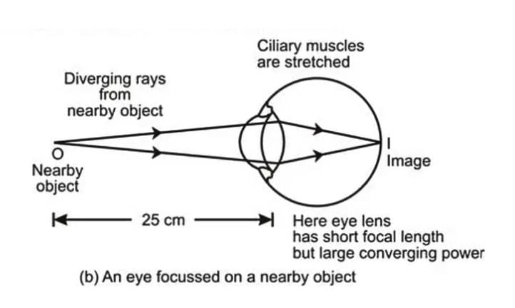
Focusing on Nearby Objects
When the eye shifts focus to a nearby object, the ciliary muscle contracts, creating tension. This causes:
A slight increase in the thickness of the eye lens.
A decrease in the focal length, allowing the image of the nearby object to form on the retina.
Clear vision of the nearby object due to this precise adjustment.
The process of adjusting the focal length happens so quickly that we are typically unaware of the changes. The ciliary muscles work seamlessly to ensure sharp vision across a range of distances.
Limit of Accommodation
The limit of accommodation refers to the range within which the eye can adjust its focal length to focus clearly on objects at different distances. A normal, healthy adult eye can accommodate objects located between:
- Infinity: The farthest distance at which the eye can focus (distant objects).
- Least distance of distinct vision: The closest distance at which the eye can focus clearly, typically about 25 cm for a healthy adult.
Persistence of Vision
Persistence of vision is the phenomenon where the human eye retains an image (or the sensation caused by light from an object) for approximately 1/16th of a second after the object is no longer visible or has been removed.
- The image formed on the retina does not fade instantly but lingers briefly, allowing the brain to perceive continuity in visual stimuli.
- This ability of the eye to retain visual impressions enables smooth perception of motion in certain contexts.
Application in Cinematography
The persistence of vision is fundamental to motion-picture projection (cinematography):
- A movie camera records a sequence of still images on film.
- During projection, these images are displayed on a screen at a rate of approximately 24 frames per second.
- Due to persistence of vision, the brain merges these rapidly displayed images into a continuous sequence, creating the illusion of smooth, uninterrupted motion.
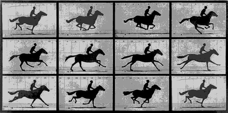
This phenomenon allows us to perceive moving pictures in films and animations as fluid and lifelike.
Colour Vision: How We See Colours
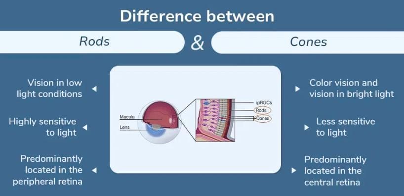
We perceive objects when their images form on the retina, the light-sensitive layer at the back of the eye. The retina contains two types of light-sensitive cells: rods and cones, which play distinct roles in vision.
Roles of Rods and Cones
- Rod-shaped cells: These cells are sensitive to the intensity of light, detecting levels of brightness or darkness. They enable vision in low-light conditions but do not contribute to colour perception.
- Cone-shaped cells: These cells are responsible for colour vision. They are sensitive to specific wavelengths of light, allowing us to distinguish between different colours.The cone-shaped cells are inactive in dim light, which is why we struggle to see colours in low-light conditions.
Differences in Colour Perception Among Animals and Birds
The structure and quantity of rod-shaped and cone-shaped cells vary across different animals and birds, leading to differences in their colour perception. For instance:
- Bees possess cones that are sensitive to ultraviolet light, allowing them to see the ultraviolet rays present in sunlight.
- Humans cannot perceive ultraviolet light because their cones are not sensitive to it.
- Chickens have a higher ratio of cone-shaped cells compared to rod-shaped cells, which limits their vision in low light. This is why they can only see well in bright light and are more active during the day.
The ability of the eye to focus on both near and distant objects by adjusting its focal length is known as the accommodation of the eye.
The closest distance at which the eye can see objects clearly without strain is termed the near point or the least distance of distinct vision.
The acronym VIBGYOR can help you remember the sequence of colours.
|
80 videos|569 docs|80 tests
|
FAQs on The Human Eye - Science Class 10
| 1. What is the structure of the human eye? |  |
| 2. How does the human eye focus on objects at different distances? | |
| 3. How does the retina send visual information to the brain? | |
| 4. What is the role of the iris in the human eye? | |
| 5. What are the common vision problems associated with the human eye? | |


The Human Eye | Science Class 10
,MCQs
,Exam
,Free
,The Human Eye | Science Class 10
,shortcuts and tricks
,The Human Eye | Science Class 10
,Objective type Questions
,Extra Questions
,Important questions
,mock tests for examination
,Sample Paper
,past year papers
,ppt
,video lectures
,study material
,Semester Notes
,practice quizzes
,Previous Year Questions with Solutions
,Viva Questions
,Summary
;


The Human Eye Free PDF Download
Importance of The Human Eye
The Human Eye Notes
The Human Eye Class 10 Questions
Study The Human Eye on the App
|
© EduRev
|
Education Revolution
|


|


