


All Exams >
NEET >
1 Year Dropper's Course for NEET >
All Questions
All questions of Neural Control and Coordination for NEET Exam
If the corpus callosum is removed in mammalian brain then what will be affected :-- a)Coordination of Cerebrum
- b)Involuntary activity of brain
- c)Coordination of Cerebellum
- d)Behaviour and emotional disturbances
Correct answer is option 'A'. Can you explain this answer?
If the corpus callosum is removed in mammalian brain then what will be affected :-
a)
Coordination of Cerebrum
b)
Involuntary activity of brain
c)
Coordination of Cerebellum
d)
Behaviour and emotional disturbances
|
|
Kadambala Hemalatha answered |
Option A is correct becose, corpus callosam is trancverce connection between two cerebral hemisphears.. it helps in coordination between the two cerebral hemisphears..

The most abundant intracellular cation is : [NEET 2013]- a)Ca++
- b)H+
- c)K+
- d)Na+
Correct answer is option 'C'. Can you explain this answer?
The most abundant intracellular cation is : [NEET 2013]
a)
Ca++
b)
H+
c)
K+
d)
Na+

|
Shalini Saha answered |
The most abundant intracellular cation is K+.
Brain stem is the support system of brain and is the collective name for :
- a)Medulla, Pons,and Midbrain
- b)Hypothalamus, Diencephalon, Cerebellum, and Pons
- c)Cerebrum, Mesencephalon, Diencephalon, and Medulla
- d)Prosencephalon, Mesencephalon, Pons, and Medulla
Correct answer is option 'A'. Can you explain this answer?
Brain stem is the support system of brain and is the collective name for :
a)
Medulla, Pons,and Midbrain
b)
Hypothalamus, Diencephalon, Cerebellum, and Pons
c)
Cerebrum, Mesencephalon, Diencephalon, and Medulla
d)
Prosencephalon, Mesencephalon, Pons, and Medulla
|
|
Pritam Choudhary answered |
Brainstem: The Support System of Brain
The brainstem is the lower part of the brain that connects the brain to the spinal cord. It is responsible for regulating important functions such as breathing, heart rate, blood pressure, and consciousness. The brainstem consists of three main parts, which are:
Medulla
The medulla oblongata, or simply the medulla, is the lowest part of the brainstem. It controls involuntary functions such as breathing, heartbeat, and digestion. The medulla also contains reflex centers for coughing, sneezing, and vomiting.
Pons
The pons is the middle part of the brainstem, located above the medulla and below the midbrain. It is involved in the regulation of sleep, respiration, and posture. The pons also helps to relay messages between different parts of the brain.
Midbrain
The midbrain, also known as the mesencephalon, is the uppermost part of the brainstem. It is involved in the regulation of vision, hearing, and movement. The midbrain contains several important structures, including the substantia nigra, which produces dopamine and is involved in the control of movement.
Conclusion
In conclusion, the brainstem is the support system of the brain and is responsible for regulating vital functions. It consists of three main parts, which are the medulla, pons, and midbrain. The medulla controls involuntary functions such as breathing and heartbeat, the pons is involved in the regulation of sleep and posture, and the midbrain is responsible for vision, hearing, and movement.
During the transmission of nerve impulse through a nerve fibre, the potential on the inner side of the plasma membrane has which type of electric change? [2007]- a)First positive, then negative and continue to be positive
- b)First negative, then positive and continue to be positive.
- c)First positive, then negative and again back to positive
- d)First negative, then positive and again back to negative
Correct answer is option 'D'. Can you explain this answer?
During the transmission of nerve impulse through a nerve fibre, the potential on the inner side of the plasma membrane has which type of electric change? [2007]
a)
First positive, then negative and continue to be positive
b)
First negative, then positive and continue to be positive.
c)
First positive, then negative and again back to positive
d)
First negative, then positive and again back to negative

|
Muskaan Basak answered |
Once the events of depolarization have occurred, a nerve impulse or spike is initiated. Action potential is another name of nerve impulse. It lasts about 1 msec (millisecond). The stimulalted, negatively charged point on the outside of the membrane sends out an electrical current to the positive point (still polarized adjacent to it). This local current causes the adjacent inner part of the membrane to reverse its potential from –70 mV to +30 mV. The reversal repeats itself over and over until the nerve impulse is conducted through the length of the neuron
Injury localised to the hypothalamus would most likely disrupt- a)short term memory
- b)coordination during locomotion
- c)executive function, such as decision making
- d)regulation of body temperature
Correct answer is option 'D'. Can you explain this answer?
Injury localised to the hypothalamus would most likely disrupt
a)
short term memory
b)
coordination during locomotion
c)
executive function, such as decision making
d)
regulation of body temperature

|
Aman Sharma answered |
The primary function of the hypothalamus is homoeostasis, which is to maintain the body's normal temperature and body's status. So, if there is an injury to the hypothalamus, the thermoregulatory centre is disturbed resulting in a deregulation of body temperature. So, the correct answer is option D.
What is meant by coordination?- a)Only two organs interact
- b)Two or more organs interact
- c)Only two organs systems interact
- d)Only three organs interact
Correct answer is option 'B'. Can you explain this answer?
What is meant by coordination?
a)
Only two organs interact
b)
Two or more organs interact
c)
Only two organs systems interact
d)
Only three organs interact

|
EduRev NEET answered |
Coordination is the process through which two or more organs interact and complement the functions of one another. For example, the functions of muscles, lungs, heart, blood vessels, kidney, and other organs are coordinated while performing physical exercises.
Topic in NCERT: NEURAL CONTROL AND COORDINATIONLine in NCERT: "Coordination is the process through which two or more organs interact and complement the functions of one another."
The function of an axon is :-- a)Transformation of nerve impulse
- b)Reception of stimuli from neurons
- c)Reception of external stimuli
- d)Conduction of nerve impulse
Correct answer is option 'D'. Can you explain this answer?
The function of an axon is :-
a)
Transformation of nerve impulse
b)
Reception of stimuli from neurons
c)
Reception of external stimuli
d)
Conduction of nerve impulse
|
|
Sethulakshmi E.d Dinesan answered |
Axon transmit impulses away from cellbody to a synapse or to a neuro muscular junction.(11th ncert page no 317 .5th line)
The Schwann sheath is :-- a)A non myelinated nerve fibres
- b)Associated with myelin sheath
- c)A connective tissue cell
- d)Associated with myelinated & non myelinated nerve fibre
Correct answer is option 'D'. Can you explain this answer?
The Schwann sheath is :-
a)
A non myelinated nerve fibres
b)
Associated with myelin sheath
c)
A connective tissue cell
d)
Associated with myelinated & non myelinated nerve fibre
|
|
Rohan Singh answered |
Neurilemma (also known as neurolemma, sheath of Schwann, or Schwann's sheath) is the outermost nucleated cytoplasmic layer of Schwann cells (also called neurilemmocytes) that surrounds the axon of the neuron. It forms the outermost layer of the nerve fiber in the peripheral nervous system.
The membrane which cover the brain and the spinal cord is :-
- a)White matter
- b)Grey matter
- c)Peritonium
- d)Meninges
Correct answer is option 'D'. Can you explain this answer?
The membrane which cover the brain and the spinal cord is :-
a)
White matter
b)
Grey matter
c)
Peritonium
d)
Meninges

|
Ruchi Chakraborty answered |
Meninges are the membranes that cover and protect the brain and spinal cord. There are three layers of meanings: dura mater (closest to the bone), arachnoid (loosely around the brain), pia mater (closely attached to the brain and spinal cord surface).
Which of the following is a richly vascular layer with lots of blood capillaries :-- a)Duramater
- b)Piamater
- c)Epidermis of skin
- d)Both (1) & (2)
Correct answer is option 'B'. Can you explain this answer?
Which of the following is a richly vascular layer with lots of blood capillaries :-
a)
Duramater
b)
Piamater
c)
Epidermis of skin
d)
Both (1) & (2)
|
|
Riya Banerjee answered |
The pia mater of the brain is the innermost of the three membranes, that cover it. It is the vascular membrane of the brain. It carries the minute branches of the two internal carotids and the two vertebral arteries. It also returns the blood to the heart. The dura mater is the most external membrane of the brain. It forms the internal periosteum of the skull. The dura mater is a dense, tough, inelastic fibrous membrane. The epidermis contains no blood vessels, and cells in the deepest layers are nourished by diffusion from blood capillaries extending to the upper layers of the dermis. So, the correct answer is option B.
During the propagation of a nerve impulse, the action potential results from the movement of : [2008]- a)K+ ions from extracellular fluid to intracellular fluid
- b)Na+ ions from intracellular fluid to extracellular fluid
- c)K+ ions from intracellular fluid to extracellular fluid
- d)Na+ ions from extracellular fluid to intracellular fluid
Correct answer is option 'D'. Can you explain this answer?
During the propagation of a nerve impulse, the action potential results from the movement of : [2008]
a)
K+ ions from extracellular fluid to intracellular fluid
b)
Na+ ions from intracellular fluid to extracellular fluid
c)
K+ ions from intracellular fluid to extracellular fluid
d)
Na+ ions from extracellular fluid to intracellular fluid

|
Anjana Dasgupta answered |
Total sum of physio-electrochemical changes that takes place along the length of nerve fibre is known as nerve impulse. Change in potential due to stimulation of nerve fibre is called action potential. During propagation of nerve impulse, Na⁺ enters inside so(+ve) change is formed inside the membrane. K⁺ ions come out.
Corpus callosum connects :-- a)Two cerebral hemisphere
- b)Two optic lobes
- c)Two olfactory lobes
- d)Optic chiasma
Correct answer is option 'A'. Can you explain this answer?
Corpus callosum connects :-
a)
Two cerebral hemisphere
b)
Two optic lobes
c)
Two olfactory lobes
d)
Optic chiasma
|
|
Anjali Iyer answered |
The part of the brain that connects the two hemispheres of the brain is called the corpus callosum. It contains a bundle of neuronal fibers found in humans and other higher order mammals that allow the two hemispheres to talk to one another.
During the propagation of a nerve impulse, the action potential results from the movement of- a)K+ ions from intracellular fluid to extracellular fluid
- b)Na+ ions from extracellular fluid to intracellular fluid
- c)K+ ions from extracellular fluid to intracellular fluid
- d)Na+ ions from intracellular fluid to extracellular fluid
Correct answer is option 'B'. Can you explain this answer?
During the propagation of a nerve impulse, the action potential results from the movement of
a)
K+ ions from intracellular fluid to extracellular fluid
b)
Na+ ions from extracellular fluid to intracellular fluid
c)
K+ ions from extracellular fluid to intracellular fluid
d)
Na+ ions from intracellular fluid to extracellular fluid
|
|
Vivek Patel answered |
Action potential is a change in electrical potential that occurs across a plasma membrane during the passage of a nerve impulse. During this period, there is a localized and translent switch in electric potential across the membrane from -70 mV to +45 mV. It is due to the fact that the sodium channels open and the potassium channels remain closed. As a result, sodium channels permit the influx of Na+ by diffusion from extracellular fluid to intracellular fluid.
Which of the following statements is incorrect regarding electrical synapses?- a)Transmission of signals is faster than chemical synapses
- b)Pre and postsynaptic membranes are in very close proximity
- c) Electrical synapse can flow directly from one neuron to another
- d)They are very common in our system
Correct answer is option 'D'. Can you explain this answer?
Which of the following statements is incorrect regarding electrical synapses?
a)
Transmission of signals is faster than chemical synapses
b)
Pre and postsynaptic membranes are in very close proximity
c)
Electrical synapse can flow directly from one neuron to another
d)
They are very common in our system

|
Mohit Rajpoot answered |
Electrical synapses are very rare in our system. At electrical synapses, the membranes of pre and post synaptic neurons are in very close proximity. Impulse transmission across an electrical synapse is always faster than chemical synapse.
Topic in NCERT: Transmission of ImpulsesLine in NCERT: "Electrical synapses are rare in our system."
Where are the myelinated neurons found?- a)In Spinal cord and cranial nerves
- b)Only in the embryonic condition
- c)In peripheral nerve
- d)In motor neurons
Correct answer is option 'A'. Can you explain this answer?
Where are the myelinated neurons found?
a)
In Spinal cord and cranial nerves
b)
Only in the embryonic condition
c)
In peripheral nerve
d)
In motor neurons

|
Ambition Institute answered |
Myelinated nerve fibres are found in spinal and cranial nerves. Unmyelinated nerve fibres are enclosed by a Schwann cell that does not form a myelin sheath around the axon and is commonly found in autonomous and the somatic neural system.
The rise in stimulus-induced permeability to
a. Potassium ions
b. Sodium ions
c. Restoring RMP
d. Diffusion of potassium ionsArrange them in order.- a)b-a-c-d
- b)a-b-c-d
- c)b-a-d-c
- d)c-a-d-b
Correct answer is option 'C'. Can you explain this answer?
The rise in stimulus-induced permeability to
a. Potassium ions
b. Sodium ions
c. Restoring RMP
d. Diffusion of potassium ions
a. Potassium ions
b. Sodium ions
c. Restoring RMP
d. Diffusion of potassium ions
Arrange them in order.
a)
b-a-c-d
b)
a-b-c-d
c)
b-a-d-c
d)
c-a-d-b
|
|
Ameya Khanna answered |
Understanding the Sequence of Events
In the context of neuronal action potentials and membrane permeability, the sequence of events following a stimulus is crucial. Here's the breakdown of the correct order:
1. Rise in Stimulus-Induced Permeability
- The initial step involves a rise in membrane permeability to specific ions.
- This permeability change is often triggered by a stimulus, such as neurotransmitter release or mechanical stimulation.
2. Diffusion of Potassium Ions
- After the permeability to sodium ions increases, potassium ion channels may also open.
- Potassium ions will begin to diffuse out of the cell, contributing to the repolarization phase of the action potential.
3. Restoring Resting Membrane Potential (RMP)
- As potassium ions exit the neuron, the membrane potential begins to return to its resting state.
- This restoration occurs as the cell becomes less positive inside, eventually stabilizing at the RMP.
4. Rise in Permeability to Sodium Ions
- Initially, the permeability rises for sodium ions, allowing them to enter the neuron.
- This influx of sodium causes depolarization, leading to the action potential spike.
Conclusion
- Therefore, the correct order of events is: Rise in stimulus-induced permeability (to sodium), diffusion of potassium ions, restoring RMP, and finally, the impact of sodium ion permeability.
- Hence, the correct sequence is option 'c': b-a-d-c.
Understanding this sequence is key in grasping how neurons communicate and respond to stimuli, especially during action potentials.
Assertion: The somatic neural system regulates involuntary functions of the body.
Reason: The somatic neural system is divided into the sympathetic and parasympathetic systems.- a)Both Assertion and Reason are correct, and Reason is the correct explanation of Assertion.
- b)Both Assertion and Reason are correct, but Reason is not the correct explanation of Assertion.
- c)Assertion is correct, but Reason is incorrect.
- d)Both Assertion and Reason are incorrect
Correct answer is option 'D'. Can you explain this answer?
Assertion: The somatic neural system regulates involuntary functions of the body.
Reason: The somatic neural system is divided into the sympathetic and parasympathetic systems.
Reason: The somatic neural system is divided into the sympathetic and parasympathetic systems.
a)
Both Assertion and Reason are correct, and Reason is the correct explanation of Assertion.
b)
Both Assertion and Reason are correct, but Reason is not the correct explanation of Assertion.
c)
Assertion is correct, but Reason is incorrect.
d)
Both Assertion and Reason are incorrect
|
|
Maitri Deshpande answered |
Understanding the Somatic Neural System
The assertion and reason provided in the question relate to the functions of the somatic and autonomic nervous systems.
Assertion Explained
- The somatic neural system primarily controls voluntary functions.
- It is responsible for movements of skeletal muscles, allowing conscious control over these actions.
Reason Clarification
- The somatic neural system is not divided into sympathetic and parasympathetic systems.
- Instead, these divisions belong to the autonomic nervous system, which regulates involuntary functions such as heart rate, digestion, and respiratory rate.
Correctness of Assertion and Reason
- The assertion is incorrect because it states that the somatic neural system regulates involuntary functions, which is not true.
- The reason is also incorrect because it misidentifies the components of the nervous system.
Conclusion
Given that both the assertion and reason are incorrect, the answer to the question is option 'D':
- Both Assertion and Reason are incorrect.
This understanding is crucial for students preparing for exams like NEET, as it highlights the distinctions between the somatic and autonomic nervous systems.
Read the following statements and mark the correct option which is suitable. - The neural system of animals is composed of specialized cells called neurons.
- The central neural system (CNS) consists of the brain and spinal cord.
- The peripheral nervous system (PNS) is involved in transmitting impulses only from the CNS to muscles.
- The autonomic neural system controls voluntary actions like skeletal muscle movement.
- a)Statements 1, 2, and 4 are correct.
- b)Statements 1, 2, and 3 are correct.
- c) Statements 1 and 2 are correct, while 3 and 4 are incorrect.
- d)All statements are correct.
Correct answer is option 'C'. Can you explain this answer?
Read the following statements and mark the correct option which is suitable.
- The neural system of animals is composed of specialized cells called neurons.
- The central neural system (CNS) consists of the brain and spinal cord.
- The peripheral nervous system (PNS) is involved in transmitting impulses only from the CNS to muscles.
- The autonomic neural system controls voluntary actions like skeletal muscle movement.
a)
Statements 1, 2, and 4 are correct.
b)
Statements 1, 2, and 3 are correct.
c)
Statements 1 and 2 are correct, while 3 and 4 are incorrect.
d)
All statements are correct.

|
Stepway Academy answered |
Statements 1 and 2 are correct. Statement 3 is incorrect because the PNS transmits impulses both to and from the CNS, and statement 4 is incorrect because the autonomic system controls involuntary actions.
Topic in NCERT: HUMAN NEURAL SYSTEM
Line in NCERT: "The human neural system is divided into two parts: (i) the central neural system (CNS) (ii) the peripheral neural system (PNS). The CNS includes the brain and the spinal cord and is the site of information processing and control." "The PNS is divided into two divisions called somatic neural system and autonomic neural system. The somatic neural system relays impulses from the CNS to skeletal muscles while the autonomic neural system transmits impulses from the CNS to the involuntary organs and smooth muscles of the body."
In the human neural system, the coordination between different organ systems is essential for maintaining homeostasis during physical activities.
Which of the following statements best explains the roles of the central nervous system (CNS) and the peripheral nervous system (PNS) during intense physical exercise?- a)The PNS coordinates all involuntary actions during physical exercise, including the regulation of skeletal muscles, heart rate, and respiration, while the CNS only provides feedback through the sympathetic and parasympathetic systems without direct control of the muscles
- b)The CNS exclusively controls the skeletal muscles through the somatic neural system, while the PNS regulates the automatic functions of the heart and lungs through the autonomic system, without requiring feedback to the CNS.
- c)The CNS coordinates the entire response to exercise by transmitting efferent signals to skeletal muscles through the somatic system and to involuntary organs through the autonomic system, while the PNS transmits feedback only from the muscles, excluding heart and lung responses.
- d) The CNS processes sensory information from the muscles and organs, and sends efferent impulses via the PNS to regulate heart rate and muscle contraction, while the PNS carries afferent impulses from the lungs and heart to the CNS for feedback.
Correct answer is option 'D'. Can you explain this answer?
In the human neural system, the coordination between different organ systems is essential for maintaining homeostasis during physical activities.
Which of the following statements best explains the roles of the central nervous system (CNS) and the peripheral nervous system (PNS) during intense physical exercise?
Which of the following statements best explains the roles of the central nervous system (CNS) and the peripheral nervous system (PNS) during intense physical exercise?
a)
The PNS coordinates all involuntary actions during physical exercise, including the regulation of skeletal muscles, heart rate, and respiration, while the CNS only provides feedback through the sympathetic and parasympathetic systems without direct control of the muscles
b)
The CNS exclusively controls the skeletal muscles through the somatic neural system, while the PNS regulates the automatic functions of the heart and lungs through the autonomic system, without requiring feedback to the CNS.
c)
The CNS coordinates the entire response to exercise by transmitting efferent signals to skeletal muscles through the somatic system and to involuntary organs through the autonomic system, while the PNS transmits feedback only from the muscles, excluding heart and lung responses.
d)
The CNS processes sensory information from the muscles and organs, and sends efferent impulses via the PNS to regulate heart rate and muscle contraction, while the PNS carries afferent impulses from the lungs and heart to the CNS for feedback.
|
|
Jithin Unni answered |
Understanding the Role of CNS and PNS in Physical Exercise
During intense physical exercise, the coordination between the central nervous system (CNS) and peripheral nervous system (PNS) is vital for maintaining homeostasis and ensuring efficient body function.
Functions of the CNS
- The CNS is responsible for processing sensory information from various sources, including muscles and organs.
- It generates appropriate motor responses by sending efferent impulses to skeletal muscles, facilitating movement.
- The CNS integrates feedback from the body to adjust physiological responses during exercise, ensuring that the body can meet the demands of physical activity.
Functions of the PNS
- The PNS serves as a communication network between the CNS and the rest of the body.
- It transmits afferent impulses from the organs (like the heart and lungs) back to the CNS, providing critical feedback on the body’s state during exercise.
- The PNS also carries efferent signals to involuntary organs, regulating heart rate and respiration, which are crucial for sustaining energy levels and oxygen supply during intense workouts.
Coordination for Homeostasis
- Together, the CNS and PNS work synergistically to maintain homeostasis during physical exertion.
- The CNS processes information and adjusts body functions, while the PNS ensures that feedback loops function effectively, allowing for real-time adjustments to heart rate and muscle contractions.
In summary, option D accurately describes how the CNS and PNS collaborate to manage the body’s response to exercise, ensuring that both voluntary and involuntary functions are well-coordinated for optimal performance and safety.
In the resting state of the neural membrane, diffusion due to concentration gradients, if allowed, would drive [2004]- a)K+ into the cell
- b)K+ and Na+ out of the cell
- c)Na+ into the cell
- d)Na+ out of the cell
Correct answer is option 'C'. Can you explain this answer?
In the resting state of the neural membrane, diffusion due to concentration gradients, if allowed, would drive [2004]
a)
K+ into the cell
b)
K+ and Na+ out of the cell
c)
Na+ into the cell
d)
Na+ out of the cell

|
Harshitha Dey answered |
In a resting state of the neural membrane, Na+ concentration is higher on the outer side and K+ concentration is more within the cell. This concentration gradient is maintained by voltage gated channels. Hence if diffusion is allowed Na+ would enter the cell and K+ would leave.
Where is the midbrain located?- a)Between hypothalamus and pons
- b)Between cerebrum and hypothalamus
- c)Between cerebellum and medulla
- d)Between pons and medulla
Correct answer is option 'A'. Can you explain this answer?
Where is the midbrain located?
a)
Between hypothalamus and pons
b)
Between cerebrum and hypothalamus
c)
Between cerebellum and medulla
d)
Between pons and medulla

|
Top Rankers answered |
The midbrain is located between the hypothalamus of the forebrain and the pons of the hindbrain. The midbrain is also known as the mesencephalon and controls several motor movements.
The………. difference across the resting membrane is called as Resting potential.- a)Electrochemical potential
- b)Electrical potential
- c)Chemical
- d)Chemiosmotic
Correct answer is option 'B'. Can you explain this answer?
The………. difference across the resting membrane is called as Resting potential.
a)
Electrochemical potential
b)
Electrical potential
c)
Chemical
d)
Chemiosmotic
|
|
Stepway Academy answered |
The electrical potential difference across the resting plasma membrane is called as the resting potential.
Topic in NCERT: NEURAL CONTROL AND COORDINATIONLine in NCERT: "The electrical potential difference across the resting plasma membrane is called as the resting potential."
Where are the specific receptors of neurotransmitters present?- a)Synaptic cleft
- b)Post-synaptic membrane
- c)Pre-synaptic membrane
- d)Synaptic vesicle
Correct answer is option 'B'. Can you explain this answer?
Where are the specific receptors of neurotransmitters present?
a)
Synaptic cleft
b)
Post-synaptic membrane
c)
Pre-synaptic membrane
d)
Synaptic vesicle

|
Lead Academy answered |
The released neurotransmitters bind to their specific receptors which are present on the post-synaptic membrane. The new potential developed may be either excitatory or inhibitory.
Read the given statements and select the correct ones.
(i) Autonomic neural system transmits impulses from the CNS to the voluntary organs and striated muscles of the body.
(ii) Unmyelinated nerve fibres do not have Schwann cells which form the myelin sheath.
(iii) Axonal membrane of a neuron while not conducting any impulse is comparatively more permeable to potassium ions (K+) than to sodium ions (Na+).
(iv) A synapse is formed by the membranes of a presynaptic neuron and a post synaptic neuron.- a)(i) and (ii)
- b)(ii) and (iii)
- c)(iii) and (iv)
- d)(i) and (iv)
Correct answer is option 'C'. Can you explain this answer?
Read the given statements and select the correct ones.
(i) Autonomic neural system transmits impulses from the CNS to the voluntary organs and striated muscles of the body.
(ii) Unmyelinated nerve fibres do not have Schwann cells which form the myelin sheath.
(iii) Axonal membrane of a neuron while not conducting any impulse is comparatively more permeable to potassium ions (K+) than to sodium ions (Na+).
(iv) A synapse is formed by the membranes of a presynaptic neuron and a post synaptic neuron.
a)
(i) and (ii)
b)
(ii) and (iii)
c)
(iii) and (iv)
d)
(i) and (iv)
|
|
Anjali Sharma answered |
Autonomic neural system controls and coordinates such organs which are under involuntary control and unstriated muscles of the body. Unmyelinated nerve fibres have Schwann cells which do not form myelin sheath.
Which of the following statements is/are incorrect about the electrical synapse?
(i) At electrical synapses, the membranes of pre and post synaptic neurons are in very close proximity.
(ii) Electrical current can flow directly from one neuron into the other across the synapses
(iii) Transmission of an impulse across electrical synapses is very similar to impulse conduction along single axon
(iv) Electrica synapses pass electrical signal between cells with the use of Ach.
(v) Electrical synapses are fast.
(vi) Electrical synapses are rare in our system.- a)(ii), (iv) and (v)
- b)(i) and (iii)
- c)(iv) only
- d)(i), (v) and (vi)
Correct answer is option 'C'. Can you explain this answer?
Which of the following statements is/are incorrect about the electrical synapse?
(i) At electrical synapses, the membranes of pre and post synaptic neurons are in very close proximity.
(ii) Electrical current can flow directly from one neuron into the other across the synapses
(iii) Transmission of an impulse across electrical synapses is very similar to impulse conduction along single axon
(iv) Electrica synapses pass electrical signal between cells with the use of Ach.
(v) Electrical synapses are fast.
(vi) Electrical synapses are rare in our system.
a)
(ii), (iv) and (v)
b)
(i) and (iii)
c)
(iv) only
d)
(i), (v) and (vi)
|
|
Soumya Kulkarni answered |
Incorrect Statements about Electrical Synapse:
Membranes Proximity:
- Statement (i) is incorrect because at electrical synapses, the membranes of pre and post synaptic neurons are in direct contact, not just in close proximity.
Direct Current Flow:
- Statement (ii) is correct as electrical current can flow directly from one neuron into the other across the synapses.
Impulse Transmission:
- Statement (iii) is incorrect as the transmission of an impulse across electrical synapses is not similar to impulse conduction along a single axon. It is faster and more direct.
Use of Ach:
- Statement (iv) is incorrect because electrical synapses do not pass electrical signals between cells using acetylcholine (Ach) or any other neurotransmitter.
Speed of Transmission:
- Statement (v) is correct as electrical synapses are known for their fast transmission of signals between neurons.
Prevalence of Electrical Synapses:
- Statement (vi) is incorrect as electrical synapses are actually quite common in the nervous system, especially in regions where rapid signaling is essential.
Therefore, the incorrect statements about electrical synapses are (i), (iii), and (iv), making option (c) the correct choice.
Which of the following correctly describes the role of the central nervous system (CNS)?- a) It is responsible for transmitting impulses from the organs to the brain.
- b) It processes information and controls bodily functions.
- c) It includes all the nerves outside the brain and spinal cord.
- d) It consists only of the spinal cord.
Correct answer is option 'B'. Can you explain this answer?
Which of the following correctly describes the role of the central nervous system (CNS)?
a)
It is responsible for transmitting impulses from the organs to the brain.
b)
It processes information and controls bodily functions.
c)
It includes all the nerves outside the brain and spinal cord.
d)
It consists only of the spinal cord.
|
|
Ambition Institute answered |
The central nervous system (CNS) is primarily responsible for processing information and regulating bodily functions. It encompasses the brain and spinal cord, serving as the main control center for all activities in the body. The CNS interprets sensory information received from the peripheral nervous system (PNS) and coordinates responses. An interesting fact is that the brain alone contains approximately 86 billion neurons, which play a crucial role in cognitive functions, movement, and sensory perception.
Which of the following statements is correct for ‘nodes of Ranvier’ of nerve? [2002]- a)Neurilemma is discontinuous
- b)Myelin sheath is discontinuous
- c)Both neurilemma and myelin sheath are discontinuous
- d)Covered by myelin sheath
Correct answer is option 'B'. Can you explain this answer?
Which of the following statements is correct for ‘nodes of Ranvier’ of nerve? [2002]
a)
Neurilemma is discontinuous
b)
Myelin sheath is discontinuous
c)
Both neurilemma and myelin sheath are discontinuous
d)
Covered by myelin sheath
|
|
Srestha Bose answered |
Understanding Nodes of Ranvier
The nodes of Ranvier are critical components in the structure of myelinated nerve fibers. They play a significant role in the propagation of nerve impulses through a process known as saltatory conduction.
Discontinuity of Myelin Sheath
- The myelin sheath is a protective covering that surrounds the axons of many neurons.
- At the nodes of Ranvier, this myelin sheath is interrupted.
- This discontinuity allows for the rapid transmission of electrical signals along the nerve fiber.
Role of Neurilemma
- Neurilemma, also known as the Schwann cell sheath, is present in myelinated fibers.
- While the neurilemma is found at the nodes, it is primarily the myelin sheath that is discontinuous at these points.
- Neurilemma does not contribute to the discontinuity of the myelin sheath but rather supports the axon.
Importance of Myelin Discontinuity
- The gaps created by the nodes of Ranvier facilitate the jumping of action potentials from one node to the next.
- This process significantly increases the speed of nerve impulse conduction compared to unmyelinated fibers.
Conclusion
In summary, the correct statement regarding the nodes of Ranvier is that the myelin sheath is discontinuous at these points. This structural feature is essential for efficient nerve signal transmission, making option 'B' the accurate choice.
Which part of the neuron is present in a high concentration in the grey matter?- a)Cell body
- b)Axon
- c)Dendrites
- d)Synaptic knobs
Correct answer is option 'A'. Can you explain this answer?
Which part of the neuron is present in a high concentration in the grey matter?
a)
Cell body
b)
Axon
c)
Dendrites
d)
Synaptic knobs
|
|
Gayatri Desai answered |
Grey Matter and Neurons
Grey matter is a type of neural tissue found in the brain and spinal cord. It consists of cell bodies, dendrites, and synapses, and appears grey due to the presence of unmyelinated axons and glial cells. Neurons are the functional units of the nervous system and they transmit information through electrical impulses.
Concentration of Cell Bodies in Grey Matter
The grey matter contains a high concentration of cell bodies compared to other parts of the neuron. The cell body, also known as the soma or perikaryon, is the main part of the neuron that contains the nucleus and various organelles necessary for cellular functions. It is responsible for maintaining the metabolic and structural integrity of the neuron.
Functions of Cell Bodies
The cell body plays several important roles in the neuron:
1. Protein synthesis: The cell body contains ribosomes and other organelles involved in protein synthesis. It produces proteins that are essential for the survival and functioning of the neuron.
2. Metabolism: The cell body generates energy and metabolizes various substances required for neuronal processes. It produces ATP through cellular respiration to fuel the neuron's activities.
3. Integration of information: The cell body receives signals from dendrites and processes them. It integrates these signals and determines whether to transmit or inhibit the electrical impulse.
4. Structural support: The cell body provides structural support to the neuron by giving it its shape and stability. It also connects to other neurons through synapses.
Conclusion
In conclusion, the cell body is present in a high concentration in the grey matter. It is responsible for various vital functions such as protein synthesis, metabolism, integration of information, and structural support. Understanding the distribution and functions of different parts of the neuron, including the cell body, is essential in comprehending the complexity of the nervous system and its role in various physiological processes.
During refractory period :-- a)Nerve transmits impulse very slowly
- b)Nerve can not transmit impulse
- c)Nerve transmits impulses very rapidly
- d)None of the above
Correct answer is option 'B'. Can you explain this answer?
During refractory period :-
a)
Nerve transmits impulse very slowly
b)
Nerve can not transmit impulse
c)
Nerve transmits impulses very rapidly
d)
None of the above
|
|
Rohan Singh answered |
This is the time during which another stimulus given to the neuron (no matter how strong) will not lead to a second action potential. Thus, because Na+ channels are inactivated during this time, additional depolarizing stimuli do not lead to new action potentials. The absolute refractory period takes about 1-2 ms. refractory period is a period of time during which an organ or cell is incapable of repeating a particular action, or (more precisely) the amount of time it takes for an excitable membrane to be ready for a second stimulus once it returns to its resting state following an excitation. It most commonly refers to electrically excitable muscle cells or neurons. Absolute refractory period corresponds to depolarization and repolarization, whereas relative refractory period corresponds to hyperpolarization.
When a neuron is in resting state I not conducting any impulse, the axonal membrane is: [2011]- a)comparatively more permeable to Na+ ions and nearly impermeable to K+ ions
- b)equally permeable to both Na+ and K+ ions
- c)impermeable to both Na+ and K+ ions
- d)comparatively more permeable to K+ ions and nearly impermeable to Na+ ions
Correct answer is option 'D'. Can you explain this answer?
When a neuron is in resting state I not conducting any impulse, the axonal membrane is: [2011]
a)
comparatively more permeable to Na+ ions and nearly impermeable to K+ ions
b)
equally permeable to both Na+ and K+ ions
c)
impermeable to both Na+ and K+ ions
d)
comparatively more permeable to K+ ions and nearly impermeable to Na+ ions

|
Pallabi Reddy answered |
When a neurone is in resting state i.e., not conducting any impulse, the axonal membrane is comparatively more permeable to K+ ion and nearly impermeable to Na+ ions.
The box like bony structure which encloses the brain is called :-- a)Cranium
- b)Pericardium
- c)Peritoneum
- d)Periosteum
Correct answer is option 'A'. Can you explain this answer?
The box like bony structure which encloses the brain is called :-
a)
Cranium
b)
Pericardium
c)
Peritoneum
d)
Periosteum

|
Ramesh Chand answered |
The box enclosing and protecting the brain is called as the cranium . The cranium is the part of the skull, that encloses the brain. Therefore option A is the correct answer.
Cerebellum is concerned with :-- a)Co-ordination of muscular movement
- b)Memory
- c)Vision
- d)Reflex action
Correct answer is option 'A'. Can you explain this answer?
Cerebellum is concerned with :-
a)
Co-ordination of muscular movement
b)
Memory
c)
Vision
d)
Reflex action
|
|
Sanjana Singh answered |
Cerebellum is a part of the brain and is responsible for motor control which includes muscle movement , equilibrium and balance as it relates to movement .
So 'a' is correct.
So 'a' is correct.
Which structure connects the two hemispheres of the cerebrum?- a)Thalamus
- b)Hypothalamus
- c)Limbic system
- d) Corpus callosum
Correct answer is option 'D'. Can you explain this answer?
Which structure connects the two hemispheres of the cerebrum?
a)
Thalamus
b)
Hypothalamus
c)
Limbic system
d)
Corpus callosum
|
|
Debanshi Pillai answered |
Connection Between Hemispheres
The structure that connects the two hemispheres of the cerebrum is the Corpus Callosum. This large bundle of nerve fibers plays a crucial role in interhemispheric communication.
Function of the Corpus Callosum
- Communication: It facilitates communication between the left and right hemispheres, allowing for the integration of sensory information and coordination of motor functions.
- Transfer of Information: It enables the transfer of information from one hemisphere to the other, aiding in tasks that require cooperation between both sides of the brain.
Structure of the Corpus Callosum
- Anatomy: The corpus callosum is a C-shaped structure located beneath the cerebral cortex. It consists of over 200 million axons that connect corresponding regions of the two hemispheres.
- Regions: It can be divided into four parts: the rostrum, genu, body, and splenium, each connecting different areas of the brain.
Importance in Brain Function
- Cognitive Functions: The corpus callosum is essential for higher cognitive functions such as problem-solving, language, and spatial awareness.
- Motor Coordination: It plays a vital role in coordinating movements that involve both sides of the body, ensuring they work together seamlessly.
In summary, the corpus callosum is essential for integrating the functions of the left and right hemispheres, making it a critical structure for overall brain function and coordination.
What are the short repeatedly branched fibres called?
- a)Axon
- b)Dendrite
- c)Neurite
- d)Cell body
Correct answer is option 'B'. Can you explain this answer?
What are the short repeatedly branched fibres called?
a)
Axon
b)
Dendrite
c)
Neurite
d)
Cell body
|
|
Akash Nair answered |
Short Repeatedly Branched Fibres: Axon
An axon is a long, slender projection of a neuron that conducts electrical impulses away from the cell body, towards other neurons or target cells. It is the primary transmission line of the nervous system, responsible for transmitting and propagating action potentials.
Structure of an Axon:
- Axon Hillock: The axon hillock is the cone-shaped region of the neuron where the axon originates from the cell body. It is the site where action potentials are generated.
- Axon Proper: The axon proper is the elongated part of the axon that extends away from the cell body. It is covered by a lipid-rich insulating layer called the myelin sheath, which is produced by specialized supporting cells called Schwann cells in the peripheral nervous system, and oligodendrocytes in the central nervous system.
- Axon Terminal: At the distal end of the axon, there are fine branches called axon terminals or terminal boutons. These branches form synapses with other neurons or target cells, allowing for the transmission of signals.
Function of an Axon:
- Signal Transmission: The main function of an axon is to transmit electrical signals, known as action potentials, from the cell body to other neurons or target cells. These signals travel along the axon, facilitated by the myelin sheath and nodes of Ranvier, which allow for saltatory conduction. This rapid transmission of signals enables efficient communication within the nervous system.
- Synaptic Transmission: At the axon terminals, the electrical signals are converted into chemical signals. When an action potential reaches the axon terminal, it triggers the release of neurotransmitters into the synaptic cleft, which then bind to receptors on the target cell, transmitting the signal from one neuron to another or to an effector cell (such as a muscle cell or gland).
Conclusion:
In summary, the short repeatedly branched fibers in neurons are called axons. Axons play a vital role in the transmission of electrical signals throughout the nervous system, allowing for communication between neurons and the activation of target cells.
Read the given statements and select the correct option.
(i) Synaptic cleft of neurons secrete adrenaline.
(ii) Myelinated nerve fibres are enveloped with Schwann cells, which from a myelin sheath around the axon.
(iii) Non-myelinated nerve fibre is enclosed by a Schwann cell that does not form a myelin sheath.
(iv) Spinal and cranial nerves are made of non-melinated nerve fibres.- a)Statements (i) and (ii) are correct but statements (iii) and (iv) are incorrect
- b)Statements (i), (ii) and (iii) are correct but statement (iv) is incorrect
- c)Statement (iii) and (iv) are correct but statement (i) and (ii) are incorrect
- d)Statement (ii) and (iii) are correct but statements (i) and (iv) are incorrect
Correct answer is option 'D'. Can you explain this answer?
Read the given statements and select the correct option.
(i) Synaptic cleft of neurons secrete adrenaline.
(ii) Myelinated nerve fibres are enveloped with Schwann cells, which from a myelin sheath around the axon.
(iii) Non-myelinated nerve fibre is enclosed by a Schwann cell that does not form a myelin sheath.
(iv) Spinal and cranial nerves are made of non-melinated nerve fibres.
a)
Statements (i) and (ii) are correct but statements (iii) and (iv) are incorrect
b)
Statements (i), (ii) and (iii) are correct but statement (iv) is incorrect
c)
Statement (iii) and (iv) are correct but statement (i) and (ii) are incorrect
d)
Statement (ii) and (iii) are correct but statements (i) and (iv) are incorrect
|
|
Preeti Iyer answered |
Synaptic vesicles of the synaptic knob secrete neurotransmitter (e.g., adrenaline). Spinal nerves and cranial nerves are myelinated nerves.
Which of the following statements are correct?
(A) Depolarization of an axonal membrane is caused due to rise in stimulus-induced permeability to Na⁺ and its rapid influx into axoplasm.
(B) Diffusion of K⁺ outside the axonal membrane restores the resting potential of the membrane.
(C) Sodium-potassium pump maintains active transport of 2 Na⁺ outwards for 3 K⁺ into the axoplasm across the resting membrane.- a)A only
- b)A and B
- c)B and C
- d)A and C
Correct answer is option 'B'. Can you explain this answer?
(A) Depolarization of an axonal membrane is caused due to rise in stimulus-induced permeability to Na⁺ and its rapid influx into axoplasm.
(B) Diffusion of K⁺ outside the axonal membrane restores the resting potential of the membrane.
(C) Sodium-potassium pump maintains active transport of 2 Na⁺ outwards for 3 K⁺ into the axoplasm across the resting membrane.
a)
A only
b)
A and B
c)
B and C
d)
A and C

|
Bs Academy answered |
Depolarization of the axonal membrane happens due to an increase in Na⁺ permeability, which allows Na⁺ ions to rapidly enter the axoplasm, making Statement A correct. Statement B is also correct, as the diffusion of K⁺ out of the membrane restores the resting potential. Statement C is incorrect because the sodium-potassium pump moves 3 Na⁺ out for every 2 K⁺ in, not the other way around. Hence, the correct statements are A and B.Topic in NCERT: Generation and Conduction of Nerve Impulse
Line in NCERT: "The rise in the stimulus-induced permeability to Na* is extremely short-lived. It is quickly followed by a rise in permeability to K*. Within a fraction of a second, K+ diffuses outside the membrane and restores the resting potential of the membrane at the site of excitation and the fibre becomes once more responsive to further stimulation."
CNS is mostly made of [1993]- a)motor neurons and sensory neurons
- b)sensory neurons and association neurons
- c)association neurons
- d)motor neurons and association neurons
Correct answer is option 'C'. Can you explain this answer?
a)
motor neurons and sensory neurons
b)
sensory neurons and association neurons
c)
association neurons
d)
motor neurons and association neurons
|
|
Top Rankers answered |
The central nervous system (CNS) primarily consists of association neurons (interneurons), which connect sensory neurons to motor neurons and process information within the brain and spinal cord.
How do ions contribute to the resting potential of a neuron?- a)By moving randomly across the membrane
- b)By maintaining a higher concentration of sodium inside the cell
- c)By preventing any ion movement across the membrane
- d)By establishing a high concentration of potassium inside and sodium outside
Correct answer is option 'D'. Can you explain this answer?
How do ions contribute to the resting potential of a neuron?
a)
By moving randomly across the membrane
b)
By maintaining a higher concentration of sodium inside the cell
c)
By preventing any ion movement across the membrane
d)
By establishing a high concentration of potassium inside and sodium outside
|
|
Lead Academy answered |
The resting potential of a neuron is maintained by a high concentration of potassium ions inside and sodium ions outside the cell, facilitated by the sodium-potassium pump.
Topic in NCERT: Generation and Conduction of Nerve Impulse
Line in NCERT: "Consequently, the axoplasm inside the axon contains high concentration of K* and negatively charged proteins and low concentration of Na*. In contrast, the fluid outside the axon contains a low concentration of K*, a high concentration of Na* and thus form a concentration gradient."
Sodium-potassium pump transports- a)Na+ and K+ out of the neuron
- b)Na+ and K+ into the neuron
- c)Na+ into the neuron and K+ out of the neuron
- d)K+ into the neuron and Na+ out of the neuron
Correct answer is option 'D'. Can you explain this answer?
Sodium-potassium pump transports
a)
Na+ and K+ out of the neuron
b)
Na+ and K+ into the neuron
c)
Na+ into the neuron and K+ out of the neuron
d)
K+ into the neuron and Na+ out of the neuron
|
|
Gaurav Kumar answered |
Each Na+ -K+ pump expels three Na+ ions for every two K+ ions imported.
Assertion: The functions of the organs in our body must be coordinated to maintain homeostasis.
Reason: Coordination is the process where two or more organs interact and complement each other’s functions.- a)Both Assertion and Reason are correct, and Reason is the correct explanation of Assertion.
- b)Both Assertion and Reason are correct, but Reason is not the correct explanation of Assertion.
- c)Assertion is correct, but Reason is incorrect.
- d)Assertion is incorrect, but Reason is correct.
Correct answer is option 'A'. Can you explain this answer?
Assertion: The functions of the organs in our body must be coordinated to maintain homeostasis.
Reason: Coordination is the process where two or more organs interact and complement each other’s functions.
Reason: Coordination is the process where two or more organs interact and complement each other’s functions.
a)
Both Assertion and Reason are correct, and Reason is the correct explanation of Assertion.
b)
Both Assertion and Reason are correct, but Reason is not the correct explanation of Assertion.
c)
Assertion is correct, but Reason is incorrect.
d)
Assertion is incorrect, but Reason is correct.
|
|
Nidhi Chauhan answered |
Understanding Homeostasis
Homeostasis is the process by which living organisms maintain a stable internal environment despite external changes. This involves the regulation of various physiological parameters such as temperature, pH, and electrolyte balance.
Assertion Explanation
- The assertion states that the functions of the organs in our body must be coordinated to maintain homeostasis.
- This is correct because organs do not function in isolation. For example, the respiratory system works with the circulatory system to ensure that oxygen is delivered to tissues and carbon dioxide is removed.
Reason Explanation
- The reason provided indicates that coordination is the interaction and complementing functions of two or more organs.
- This is also a correct statement as organs often work together to perform complex tasks that no single organ could achieve alone. For example, the kidneys and the endocrine system work together to regulate blood pressure and fluid balance.
Linking Assertion and Reason
- The reason directly explains the assertion by highlighting how organs complement each other to maintain homeostasis.
- For instance, during exercise, the heart rate increases (cardiovascular system), while the lungs facilitate increased oxygen intake (respiratory system), showcasing their coordinated efforts to maintain stable conditions in the body.
Conclusion
- Since both the assertion and the reason are true, and the reason correctly explains the assertion, the correct answer is option 'A': Both Assertion and Reason are correct, and Reason is the correct explanation of Assertion.
Given below are two statements: (NEET 2024)
Statement I: The cerebral hemispheres are connected by nerve tract known as corpus callosum.
Statement II: The brain stem consists of the medulla oblongata, pons and cerebrum.
In the light of the above statements, choose the most appropriate answer from the options given below:- a)Both Statement I and Statement II are correct.
- b)Both Statement I and Statement II are incorrect.
- c)Statement I is correct but Statement II is incorrect.
- d)Statement I is incorrect but Statement II is correct.
Correct answer is option 'C'. Can you explain this answer?
Given below are two statements: (NEET 2024)
Statement I: The cerebral hemispheres are connected by nerve tract known as corpus callosum.
Statement II: The brain stem consists of the medulla oblongata, pons and cerebrum.
In the light of the above statements, choose the most appropriate answer from the options given below:
Statement I: The cerebral hemispheres are connected by nerve tract known as corpus callosum.
Statement II: The brain stem consists of the medulla oblongata, pons and cerebrum.
In the light of the above statements, choose the most appropriate answer from the options given below:
a)
Both Statement I and Statement II are correct.
b)
Both Statement I and Statement II are incorrect.
c)
Statement I is correct but Statement II is incorrect.
d)
Statement I is incorrect but Statement II is correct.
|
|
Sagar Choudhary answered |
Explanation of Statement I
- Statement I: The cerebral hemispheres are connected by a nerve tract known as corpus callosum.
- This statement is correct. The corpus callosum is a thick band of nerve fibers that connects the left and right cerebral hemispheres, facilitating communication between them.
Explanation of Statement II
- Statement II: The brain stem consists of the medulla oblongata, pons, and cerebrum.
- This statement is incorrect. The brain stem is made up of the medulla oblongata, pons, and midbrain, not the cerebrum. The cerebrum is actually a separate structure that is responsible for higher brain functions, including thought and action.
Conclusion
- Given the evaluations of both statements:
- Statement I is correct.
- Statement II is incorrect.
Thus, the correct answer is option C: Statement I is correct but Statement II is incorrect.
In which animal nerve cell is present but brain is absent? [2002]- a)Sponge
- b)Earthworm
- c)Cockroach
- d)Hydra
Correct answer is option 'D'. Can you explain this answer?
In which animal nerve cell is present but brain is absent? [2002]
a)
Sponge
b)
Earthworm
c)
Cockroach
d)
Hydra
|
|
Shalini Saha answered |
A primitive type of nervous system, without brain is found in the form of intra epidermal nerve net. However, there is no brain in Hydra to coordinate the response.
A sagittal section of human-brain is shown here. Identify at least two labels from A-D. [NEET Kar. 2013]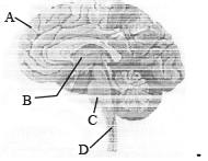
- a)A – Cerebral hemispheres; B – Cerebellum
- b)C – Mid brain; D – Cerebellum
- c)A – Cerebrum; C – Pons
- d)B – Corpus callosum; D – Medulla
Correct answer is option 'C'. Can you explain this answer?
A sagittal section of human-brain is shown here. Identify at least two labels from A-D. [NEET Kar. 2013]
a)
A – Cerebral hemispheres; B – Cerebellum
b)
C – Mid brain; D – Cerebellum
c)
A – Cerebrum; C – Pons
d)
B – Corpus callosum; D – Medulla

|
Aashna Mukherjee answered |
Cerebrum is the first and most developed part of fore brain. It makes 2/3 part of total brain. Pons is a small spherical projection, which is situated below the midbrain and upper side of the medulla oblongata. It acts as a relay centre among different parts of brain. B and D are thalamus and spinal cord respectively.
Afferent nerve fibres carry impulses from [1992]- a)effector organs to CNS
- b)receptors to CNS
- c)CNS to receptors
- d)CNS to muscles
Correct answer is option 'B'. Can you explain this answer?
a)
effector organs to CNS
b)
receptors to CNS
c)
CNS to receptors
d)
CNS to muscles
|
|
Sakshi Sharma answered |
Afferent Nerve Fibres Overview
Afferent nerve fibres play a crucial role in the functioning of the nervous system. They are responsible for transmitting sensory information from various parts of the body to the Central Nervous System (CNS).
Function of Afferent Nerve Fibres
- Direction of Impulse: Afferent fibres carry impulses from receptors located in sensory organs (like skin, eyes, ears, etc.) directly to the brain and spinal cord, which are components of the CNS.
- Sensory Information Processing: This information allows the CNS to process sensory input, enabling perception, reflexes, and appropriate responses to environmental stimuli.
Contrast with Efferent Nerve Fibres
- Efferent Nerve Fibres: Unlike afferent fibres, efferent fibres transmit signals from the CNS to effector organs, such as muscles and glands, facilitating action and response.
Importance in the Nervous System
- Homeostasis and Response: Afferent nerve fibres are essential for maintaining homeostasis as they provide critical feedback about the body’s internal and external environments.
- Reflex Actions: In reflex actions, afferent fibres quickly send signals to the CNS, which then generates a response through efferent fibres, often without conscious thought.
In summary, the correct answer to the question is option 'B', as afferent nerve fibres specifically carry impulses from receptors to the CNS, which is pivotal for sensory perception and response mechanisms within the body.
How does the pre-synaptic neuron transmit an impulse across the synaptic cleft to the post-synaptic neuron?- a)By electrical current flowing directly between the neurons across the synaptic cleft.
- b)By the release of neurotransmitters from synaptic vesicles into the synaptic cleft.
- c)By ion flow through the synaptic cleft without the need for neurotransmitters.
- d)By a direct connection between the axon terminal and the post-synaptic membrane.
Correct answer is option 'B'. Can you explain this answer?
a)
By electrical current flowing directly between the neurons across the synaptic cleft.
b)
By the release of neurotransmitters from synaptic vesicles into the synaptic cleft.
c)
By ion flow through the synaptic cleft without the need for neurotransmitters.
d)
By a direct connection between the axon terminal and the post-synaptic membrane.
|
|
Ambition Institute answered |
The pre-synaptic neuron transmits an impulse to the post-synaptic neuron by releasing neurotransmitters from synaptic vesicles into the synaptic cleft, where they bind to specific receptors on the post-synaptic membrane.
Which of the following integrates all the activities of the organs?- a)Excretory and Respiratory system
- b)The neural and digestive system
- c)Digestive and excretory system
- d)The neural and endocrine system
Correct answer is option 'D'. Can you explain this answer?
Which of the following integrates all the activities of the organs?
a)
Excretory and Respiratory system
b)
The neural and digestive system
c)
Digestive and excretory system
d)
The neural and endocrine system

|
Flembe Academy answered |
In our body, the neural and the endocrine system jointly coordinate and integrate all the activities of the organs so that they function in a synchronised fashion.
Topic in NCERT: NEURAL CONTROL AND COORDINATIONLine in NCERT: "The neural system and the endocrine system jointly coordinate and integrate all the activities of the organs so that they function in a synchronised fashion."
In Pavlov’s conditioned reflex experiment find the correct matches :
- a)a)-i, b)-iii, c)-iv, d)-ii
- b)a)-iv, b)-ii, c)-i, d)-iii
- c)a)-iii, b)-iv, c)-ii, d)-i
- d)a)-ii, b)-iii, c)-i, d)-iv
Correct answer is option 'C'. Can you explain this answer?
In Pavlov’s conditioned reflex experiment find the correct matches :
a)
a)-i, b)-iii, c)-iv, d)-ii
b)
a)-iv, b)-ii, c)-i, d)-iii
c)
a)-iii, b)-iv, c)-ii, d)-i
d)
a)-ii, b)-iii, c)-i, d)-iv
|
|
Wahid Khan answered |
There are different kinds of stimulus that is received by different receptors in our body. Hearing of sound of bell is conditioned stimulus and salivation in response to bell is conditioned response. Food itself is conditioned stimulus and salivation in response to food is unconditioned response.
Match the following
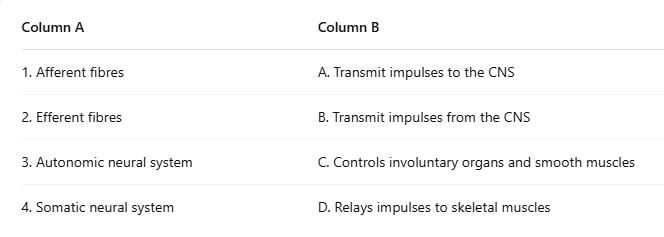
- a)1-A, 2-B, 3-D, 4-C
- b)1-B, 2-A, 3-D, 4-C
- c) 1-A, 2-B, 3-C, 4-D
- d)1-B, 2-A, 3-C, 4-D
Correct answer is option 'C'. Can you explain this answer?
Match the following
a)
1-A, 2-B, 3-D, 4-C
b)
1-B, 2-A, 3-D, 4-C
c)
1-A, 2-B, 3-C, 4-D
d)
1-B, 2-A, 3-C, 4-D
|
|
Bs Academy answered |
Afferent fibres transmit impulses to the CNS.
Efferent fibres transmit impulses from the CNS.
The autonomic neural system controls involuntary organs and smooth muscles.
The somatic neural system relays impulses to skeletal muscles.
Efferent fibres transmit impulses from the CNS.
The autonomic neural system controls involuntary organs and smooth muscles.
The somatic neural system relays impulses to skeletal muscles.
A diagram showing axon terminal and synapse is given. Identify correctly at least two of A-D. [NEET 2013]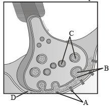
- a)B-Synaptic connection, D-K+
- b)A-Neurotransmitter, B-Synaptic cleft
- c)C- Neurotransmitter, D-Ca++
- d)A-Receptor, C-Synaptic vesicles
Correct answer is option 'D'. Can you explain this answer?
A diagram showing axon terminal and synapse is given. Identify correctly at least two of A-D. [NEET 2013]
a)
B-Synaptic connection, D-K+
b)
A-Neurotransmitter, B-Synaptic cleft
c)
C- Neurotransmitter, D-Ca++
d)
A-Receptor, C-Synaptic vesicles

|
Priyanka Iyer answered |
A-Receptor, C-Synaptic vesicles B is synaptic cleft. A synapse is formed by the membranes of a synaptic neuron and post synaptic neuron, which may or may not separated by a gap called synaptic cleft. It is filled by fluid called neurotransmitter which are involved in transmission of impulse at these synapses.
Chapter doubts & questions for Neural Control and Coordination - 1 Year Dropper's Course for NEET 2025 is part of NEET exam preparation. The chapters have been prepared according to the NEET exam syllabus. The Chapter doubts & questions, notes, tests & MCQs are made for NEET 2025 Exam. Find important definitions, questions, notes, meanings, examples, exercises, MCQs and online tests here.
Chapter doubts & questions of Neural Control and Coordination - 1 Year Dropper's Course for NEET in English & Hindi are available as part of NEET exam.
Download more important topics, notes, lectures and mock test series for NEET Exam by signing up for free.
1 Year Dropper's Course for NEET
503 videos|1699 docs|628 tests
|
|
© EduRev
|
Education Revolution
|


|


Signup to see your scores
go up
within 7 days!
within 7 days!
Takes less than 10 seconds to signup