Class 10 Exam > Class 10 Questions > Needed a Document for human eye? Related: D...
Start Learning for Free
Needed a Document for human eye?
Verified Answer
Needed a Document for human eye? Related: Dobereiner's Triads, Octav...
Human Eye
The eye is an important and one of the most complex sense organ that we humans are endowed with. It helps us in visualizing objects and also helps us in light perception, colour and depth perception. Besides, these sense organs are pretty much similar to cameras and they help us see objects when light coming from outside enters into them. That being said, it is quite interesting to understand the structure and working of a human eye. It helps us also in understanding how a camera also actually functions. Let’s have a glance on the human eye – it’s structure and function.
Structure of Human Eye
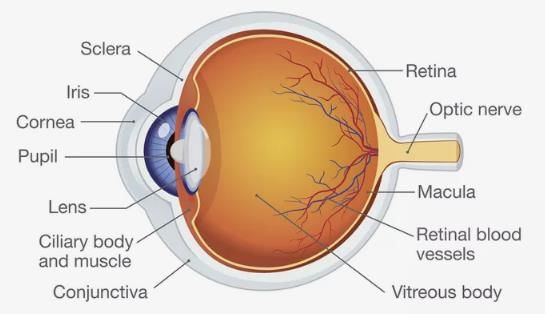
A human eye is roughly 2.3 cm in diameter and is almost a spherical ball filled with some fluid. It consists of the following parts:

Sclera: It is the outer covering, a protective tough white layer called the sclera (white part of the eye).
Cornea: The front transparent part of the sclera is called cornea. Light enters the eye through the cornea.
Iris: A dark muscular tissue and ring-like structure behind the cornea are known as the iris. The colour of iris actually indicates the colour of the eye. The iris also helps regulate or adjust exposure by adjusting the iris.
Pupil: A small opening in the iris is known as a pupil. Its size is controlled by the help of iris. It controls the amount of light that enters the eye.
Lens: Behind the pupil, there is a transparent structure called a lens. By the action of ciliary muscles, it changes its shape to focus light on the retina. It becomes thinner to focus distant objects and becomes thicker to focus nearby objects.
Retina: It is a light-sensitive layer that consists of numerous nerve cells. It converts images formed by the lens into electrical impulses. These electrical impulses are then transmitted to the brain through optic nerves.
Optic nerves: Optic nerves are of two types. These include cones and rods.
1. Cones: Cones are the nerve cells that are more sensitive to bright light. They help in detailed central and colour vision.
2. Rods: Rods are the optic nerve cells that are more sensitive to dim lights. They help in peripheral vision.
At the junction of the optic nerve and retina, there are no sensory nerve cells. So no vision is possible at that point and is known as a blind spot.
An eye also consists of six muscles. It includes the medial rectus, lateral rectus, superior rectus, inferior rectus, inferior oblique, and superior oblique. The basic function of these muscles is to provide different tensions and torques that further control the movement of the eye.
Function of the Human Eye
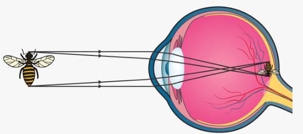
As we mentioned earlier that the eye of a human being is like a camera. Much like the electronic device, the human eye also focuses and lets in light to produce images. So basically, light rays that are deflected from or by distant objects land on the retina after they pass through various mediums like the cornea, crystalline lens, aqueous humour, the lens, and vitreous humour.

The concept here though is that as the light rays move through the various mediums, they experience refraction of light. Well, to put it in simple terms, refraction is nothing but the change in direction of the rays of light as they pass between different mediums. The table below shows the refractive indices of the various parts of the eye.
Having different refractive indexes is what bends the rays to form an image. The light rays finally are received and focused on the retina. The retina contains photoreceptor cells called rods and cones and these basically detect the intensity and the frequency of the light. Further, the image that is formed is processed by millions of these cells and they also relay the signal or nerve impulses to the brain via the optic nerve. The image formed is usually inverted but the brain corrects this phenomenon. This process is also similar to that of a convex lens.
 This question is part of UPSC exam. View all Class 10 courses
Most Upvoted Answer
Needed a Document for human eye? Related: Dobereiner's Triads, Octav...
Good.
Attention Class 10 Students!
To make sure you are not studying endlessly, EduRev has designed Class 10 study material, with Structured Courses, Videos, & Test Series. Plus get personalized analysis, doubt solving and improvement plans to achieve a great score in Class 10.

|
Explore Courses for Class 10 exam
|

|
Needed a Document for human eye? Related: Dobereiner's Triads, Octaves and Mendeleev's Periodic Table?
Question Description
Needed a Document for human eye? Related: Dobereiner's Triads, Octaves and Mendeleev's Periodic Table? for Class 10 2024 is part of Class 10 preparation. The Question and answers have been prepared according to the Class 10 exam syllabus. Information about Needed a Document for human eye? Related: Dobereiner's Triads, Octaves and Mendeleev's Periodic Table? covers all topics & solutions for Class 10 2024 Exam. Find important definitions, questions, meanings, examples, exercises and tests below for Needed a Document for human eye? Related: Dobereiner's Triads, Octaves and Mendeleev's Periodic Table?.
Needed a Document for human eye? Related: Dobereiner's Triads, Octaves and Mendeleev's Periodic Table? for Class 10 2024 is part of Class 10 preparation. The Question and answers have been prepared according to the Class 10 exam syllabus. Information about Needed a Document for human eye? Related: Dobereiner's Triads, Octaves and Mendeleev's Periodic Table? covers all topics & solutions for Class 10 2024 Exam. Find important definitions, questions, meanings, examples, exercises and tests below for Needed a Document for human eye? Related: Dobereiner's Triads, Octaves and Mendeleev's Periodic Table?.
Solutions for Needed a Document for human eye? Related: Dobereiner's Triads, Octaves and Mendeleev's Periodic Table? in English & in Hindi are available as part of our courses for Class 10.
Download more important topics, notes, lectures and mock test series for Class 10 Exam by signing up for free.
Here you can find the meaning of Needed a Document for human eye? Related: Dobereiner's Triads, Octaves and Mendeleev's Periodic Table? defined & explained in the simplest way possible. Besides giving the explanation of
Needed a Document for human eye? Related: Dobereiner's Triads, Octaves and Mendeleev's Periodic Table?, a detailed solution for Needed a Document for human eye? Related: Dobereiner's Triads, Octaves and Mendeleev's Periodic Table? has been provided alongside types of Needed a Document for human eye? Related: Dobereiner's Triads, Octaves and Mendeleev's Periodic Table? theory, EduRev gives you an
ample number of questions to practice Needed a Document for human eye? Related: Dobereiner's Triads, Octaves and Mendeleev's Periodic Table? tests, examples and also practice Class 10 tests.

|
Explore Courses for Class 10 exam
|

|
Suggested Free Tests
Signup for Free!
Signup to see your scores go up within 7 days! Learn & Practice with 1000+ FREE Notes, Videos & Tests.


